Amna Iftikhar, MD1, Nisma Faridi, MD2, Asad Jehangir, MD3, Alex Ulitsky, MD4, Daniel Rowan, MD5, Humberto Sifuentes, MD6 1Rawalpindi Medical University, Rawalpindi, Islamabad, Pakistan; 2Army Medical College, Rawalpindi, Islamabad, Pakistan; 3GI Associates LLC, Ascension Hospital, Milwaukee, WI; 4GI Associates, Milwaukee, WI; 5Ascension Hospital, Franklin, WI; 6Augusta University, Augusta, GA Introduction: Gastric glomus tumors are rare, benign mesenchymal tumors. Though often asymptomatic, patients with these tumors can also present with significant epigastric pain. Due to risk of malignant transformation, glomus tumor should be considered in the differential diagnosis of subepithelial lesions found on upper endoscopy.
Case Description/
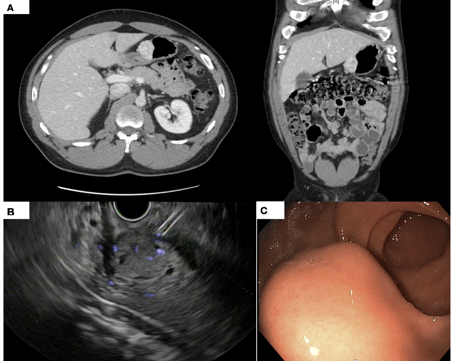
Methods: A 38-year-old African American male with no significant medical history presented with a two-month history of severe, diffuse abdominal pain, predominantly in the epigastric region. The pain, described as "excruciating", was associated with nausea and constipation. He reported 2-3 bowel movements per week despite drinking plenty of water and taking a high fiber diet. He also endorsed occasional rectal bleeding but denied unintentional weight loss. Abdominal X-ray showed moderate stool burden. The patient was advised to take polyethylene glycol. Due to persistent abdominal pain, he underwent bidirectional endoscopy. Colonoscopy showed medium sized internal hemorrhoids, but was otherwise normal. EGD revealed a 3-4 cm subepithelial lesion along the greater curvature of the stomach (Figure 1A). Abdominal CT scan showed a hyper-vascular mass, suspicious for a gastrointestinal stromal tumor or neuroendocrine tumor (Figure 1B). Endoscopic ultrasound showed a hypoechoic lesion, originating from the muscularis propria (Figure 1C). Fine-needle aspiration confirmed the diagnosis of a glomus tumor with histomorphology demonstrating epithelioid cells and immunostain positive for DOG1, synaptophysin, SMA, Calponin. The patient underwent laparoscopic partial gastrectomy, and the tumor was successfully resected. His postoperative course was uneventful, and he advanced to a soft diet on postoperative day 3. On follow up, he reported significant improvement in abdominal pain. Discussion: Gastric glomus tumors are a rare but important diagnostic consideration in patients with severe epigastric abdominal pain. Surgical resection remains the mainstay of treatment. While these tumors are typically benign, careful pathological evaluation is essential due to the possibility of malignant transformation. Long-term surveillance is recommended due to risk of recurrence.
Figure: Figure 1(A): CT scan revealing anterior gastric body mural 3.5 cm hyperattenuating round solid lesion.Figure (B).: Endoscopic ultrasound showing a hypoechoic lesion in the distal gastric body originating from muscularis propria measuring 3x3.2 cm, with small necrotic areas. The lesion abutted the left lobe of the liver without any evidence of invasion, the liver parenchyma otherwise appears normal. Figure (C).:EGD showing a subepithelial lesion in the gastric antrum along the greater curvature
Figure: Figure 2.: Gastric glomus tumor (black arrow) arising in the muscularis propria of the stomach (A, H&E, 40x). Tumor cells show uniform round nuclei surrounding hemangiopericytoma-like blood vessels with associated hyalinized stroma (B, H&E, 200x). Immunohistochemical stains show that the tumor cells are positive for SMA (C), calponin (not shown), show patchy staining with synaptophysin (D), and faint DOG1 staining (E). The tumor cells are negative for keratin AE1/AE3 (F), CD117 (G), and chromogranin (not shown).
Disclosures: Amna Iftikhar indicated no relevant financial relationships. Nisma Faridi indicated no relevant financial relationships. Asad Jehangir indicated no relevant financial relationships. Alex Ulitsky indicated no relevant financial relationships. Daniel Rowan indicated no relevant financial relationships. Humberto Sifuentes indicated no relevant financial relationships.
Amna Iftikhar, MD1, Nisma Faridi, MD2, Asad Jehangir, MD3, Alex Ulitsky, MD4, Daniel Rowan, MD5, Humberto Sifuentes, MD6. P2133 - Gastric Glomus Tumor Presenting as Severe Epigastric Pain: A Rare Case Report, ACG 2025 Annual Scientific Meeting Abstracts. Phoenix, AZ: American College of Gastroenterology.