Rhorer Legendre, MD, Jimmy Pham, MD, Vaibhav Wadhwa, MD, Sruthi Subramanian, MD McGovern Medical School at UTHealth, Houston, TX Introduction: Gastrointestinal stromal tumors (GISTs) are relatively rare, mesenchymal tumors that predominantly affect individuals between the ages of 60 and 74. They typically present on computed tomography (CT) imaging as exophytic, well-circumscribed solid masses. Here we present the case of a patient found to have a large cystic mass located between the pancreatic tail and spleen, possibly arising from the serosa of the gastric wall, that was initially thought to be a pancreatic pseudocyst but turned out to be a GIST on microscopic examination.
Case Description/
Methods: A 30-year-old man with no significant past medical history presented with epigastric and left-sided abdominal pain that started one month prior with an associated 12-pound weight loss and loss of appetite. In the emergency department, labs were notable for a normocytic anemia with hemoglobin of 10.1, compared to hemoglobin of 14.3 from an emergency department visit one month prior. CT showed a heterogeneous, enhancing, hypervascular mixed cystic and solid structure in the left upper quadrant which was in part inseparable from the greater curvature of the stomach. Endoscopic ultrasound revealed a large heterogeneous, hypoechoic abdominal mass with solid and large cystic components visualized to be originating from or touching the serosa of the gastric wall and located between the pancreatic tail and spleen. Its communication with the pancreas was unable to be visualized. The cystic component was aspirated, showing normal glucose, normal amylase, non-elevated carcinoembryonic antigen (CEA), and cytology negative for malignancy. Biopsy of the solid component revealed low-grade, spindle cell and epithelioid type GIST. The patient was subsequently referred to surgical oncology for further management. Discussion: The mass seen on endoscopic ultrasound was initially thought to be a pancreatic pseudocyst due to visualization of a large cystic component with unclear boundaries. GISTs presenting predominantly as cystic masses are relatively rare as they usually present as solid masses. Low CEA and amylase in the fluid analysis from the cyst also created a mixed picture as cystic neoplasms typically have elevated CEA and pseudocysts typically have elevated amylase. This case highlights the importance of performing a comprehensive evaluation of an abdominal mass with a presentation that is unconventional, which includes performing endoscopic ultrasound and obtaining cystic fluid aspirates and biopsy of solid components where applicable.
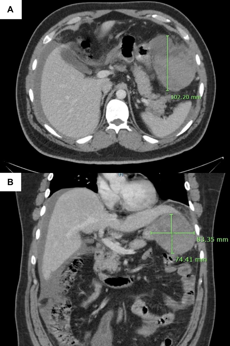
Figure: Contrast-enhanced axial (A) and coronal (B) images from a computed tomography scan of the abdomen and pelvis depicting a heterogeneous, enhancing, hypervascular mixed cystic and solid structure in the left upper quadrant measuring 10.2 x 8.3 x 7.4 cm.
Figure: Endoscopic ultrasound revealing a 9 cm by 8 cm heterogeneous, hypoechoic abdominal mass with solid and large cystic components, located between the tail of the pancreas and the spleen, with well-defined and smooth margins.
Disclosures: Rhorer Legendre indicated no relevant financial relationships. Jimmy Pham indicated no relevant financial relationships. Vaibhav Wadhwa indicated no relevant financial relationships. Sruthi Subramanian indicated no relevant financial relationships.
Rhorer Legendre, MD, Jimmy Pham, MD, Vaibhav Wadhwa, MD, Sruthi Subramanian, MD. P2126 - The Imposter Within: Unveiling the Pseudocyst Illusion, ACG 2025 Annual Scientific Meeting Abstracts. Phoenix, AZ: American College of Gastroenterology.

 photo")