Guthrie Robert Packer Hospital, Department of Internal Medicine Sayre, PA
Christopher D Manko, MD1, Abdullah Sultany, MD2, Rewanth Katamreddy, MD3, Michelle Bernshteyn, MD3, Ukorn Srivatana, MD3 1Geisinger Commonwealth School of Medicine, Sayre, PA; 2Guthrie Robert Packer Hospital, Department of Internal Medicine, Sayre, PA; 3Guthrie Robert Packer Hospital, Department of Gastroenterology, Sayre, PA Introduction: Breast cancer is the second leading cause of cancer-related death among women and commonly presents as ductal or lobular carcinoma. While metastasis to bone and brain is well known, gastric involvement is rare, particularly in the form of linitis plastica. This diffuse infiltrative pattern occurs more often with lobular than ductal carcinoma and is usually missed due to submucosal spread. Symptoms are nonspecific and may mimic chemotherapy side effects, delaying diagnosis. We report a rare case of lobular breast cancer recurrence with gastric and bony metastases presenting as linitis plastica.
Case Description/
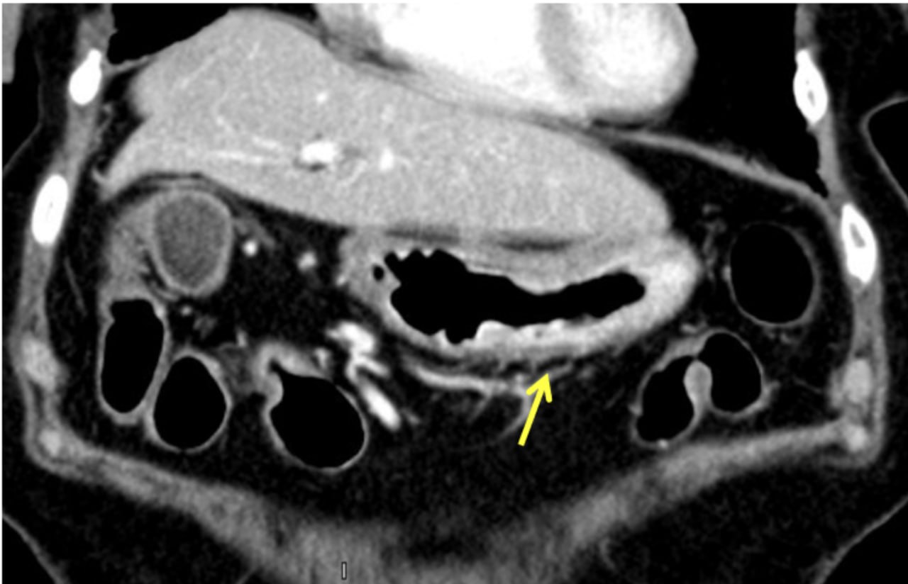
Methods: A 65-year-old woman with a history of right invasive lobular carcinoma of the breast (diagnosed in 2013, treated with mastectomy and adjuvant therapy) developed bone metastases in 2022. She was started on systemic therapy. She later developed chemotherapy-induced cardiomyopathy. In 2024, she presented with epigastric pain, nausea, early satiety, vomiting, weight loss, and belching. CT imaging revealed diffuse thickening of the gastric wall. Upper endoscopy revealed nodular gastritis; biopsies confirmed poorly differentiated lobular carcinoma (ER/PR+, E-cadherin–), consistent with gastric metastasis. Her treatment was escalated to fam-trastuzumab deruxtecan-nxki. Nine months later, she developed diarrhea initially attributed to chemotherapy. However, repeat CT showed diffuse gastric wall thickening with loss of rugal folds, consistent with linitis plastica. Symptomatic treatment resolved her diarrhea. Discussion: This case highlights a rare presentation of linitis plastica secondary to invasive lobular breast carcinoma. Given the nonspecific GI symptoms—often mistaken for chemotherapy effects—gastric metastasis may be overlooked. In our patient, persistent symptoms and CT findings of gastric wall thickening led to an endoscopic biopsy confirming metastasis. Lobular carcinoma more commonly spreads to the stomach than ductal carcinoma and often retains ER/PR positivity. Diagnosis is challenging, as linitis plastica can be submucosal and not always apparent endoscopically. A combination of imaging and targeted biopsy is often necessary. Treatment is limited to systemic therapy and palliation; prognosis remains poor, with a reported median survival of around one year. This case reinforces the importance of early recognition and a high index of suspicion in similar clinical contexts.
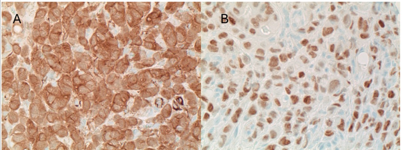
Figure: Figure 1. Histological staining of gastric biopsy with positive staining for p120 (A) and ER (B).
Disclosures: Christopher D Manko indicated no relevant financial relationships. Abdullah Sultany indicated no relevant financial relationships. Rewanth Katamreddy indicated no relevant financial relationships. Michelle Bernshteyn indicated no relevant financial relationships. Ukorn Srivatana indicated no relevant financial relationships.
Christopher D Manko, MD1, Abdullah Sultany, MD2, Rewanth Katamreddy, MD3, Michelle Bernshteyn, MD3, Ukorn Srivatana, MD3. P2119 - A Case of Invasive Lobular Carcinoma Metastasis to the Stomach, ACG 2025 Annual Scientific Meeting Abstracts. Phoenix, AZ: American College of Gastroenterology.