Bushra Khalid, MBBS, MD1, Saba Fatima, MBBS2 1Mobile Infirmary Medical Center, AL, Mobile, AL; 2King Edward Medical University, Pakistan, Lahore, Punjab, Pakistan Introduction: Gastric Trichobezoars are the foreign bodies in the stomach that are typically detected in females with psychiatric illnesses. It is a rare condition and is often considered vague and insidious in regards to its presentation until it grows into size when the symptoms start to appear.
Case Description/
Methods: A 15-year-old female patient with a 6-year history of trichophagia presented with complaints of pain in the epigastrium accompanied by nausea and off-and-on episodes of vomiting for 3 months. Examination revealed a mobile, smooth, well-defined, and hard intraabdominal mass extending from the Right Iliac region to the epigastrium and Right hypochondrium.
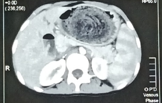
Ultrasonography showed a hypoechoic lesion subhepatically with a strong acoustic shadow. Later the upper endoscopy confirmed the diagnosis of a large well-organized trichobezoar starting just below the lower end of the esophagus and extending to the pylorus involving the fundus. Upon the CT scan of the Abdomen and Pelvis region there was a heterogenous mass of mottled gas and compressed concentric rings pattern due to the presence of entrapped air and food debris extending into the pylorus. There was a mild splenomegaly associated with this.
The mass was surgically removed via laparotomy and the post-operative period was uneventful. Discussion: Trichobezoar is usually caused by Trichophagia and Trichotillomania. Female gender, mental retardation, excessive food or nonfood substance consumption, anorexia nervosa, pica, obsessive-compulsive disorder, and depression are all factors linked to the creation of trichobezoar.
Although rare, Trichobezoar should be considered a differential diagnosis in young females who present with chronic epigastric pain and an epigastric lump. The symptoms start to appear as its size grows. In rare cases, a patient may have acute abdominal symptoms due to total intestinal obstruction or perforation.
Endoscopy and CT scans are common tools to make a diagnosis. While endoscopy shows a hair mass while CT scan can highlight other regions in addition to the stomach.
Treatment options include removal with either endoscopy, laparoscopy or laparotomy. Medical treatment consisting of various enzymes may be considered in small bezoars but is usually ineffective for large bezoars as in our case. To address the underlying emotional or physical causes and prevent recurrence, psychiatric surveillance is recommended as part of the treatment approach for patients with trichobezoars.
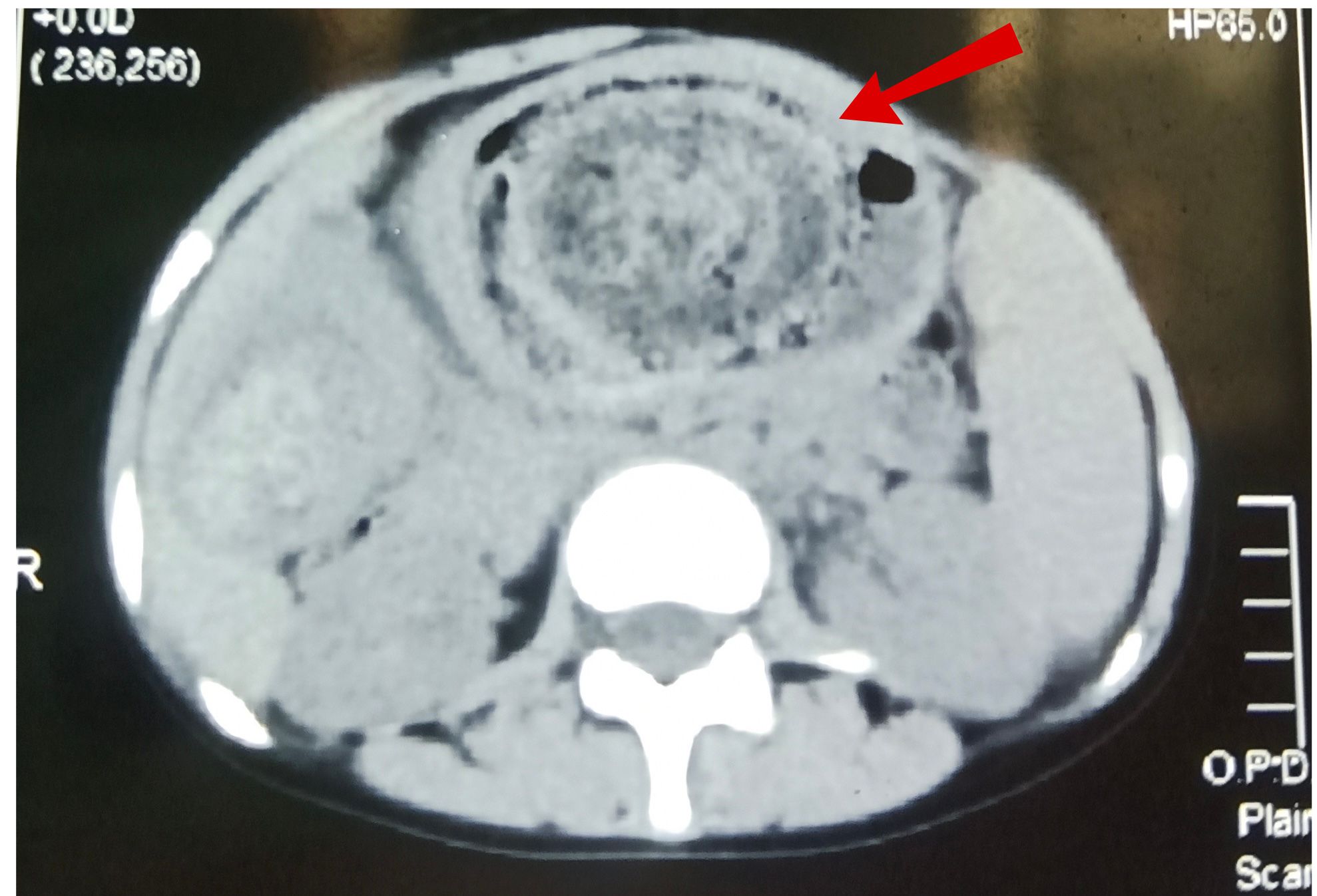
Figure: Axial plain CT imaging of the abdomen showing a large gastric trichobezoar (red arrow) with a mottled gas pattern and concentric ring appearance
Figure: Axial CT of the abdomen during the venous phase demonstrating an intragastric inhomogeneous mass with no contrast enhancement, consistent with the diagnosis of trichobezoar.
Disclosures: Bushra Khalid indicated no relevant financial relationships. Saba Fatima indicated no relevant financial relationships.
Bushra Khalid, MBBS, MD1, Saba Fatima, MBBS2. P2072 - Gastric Trichobezoar With Concentric Rings Pattern on CT: A Case Report, ACG 2025 Annual Scientific Meeting Abstracts. Phoenix, AZ: American College of Gastroenterology.