Creighton University School of Medicine Phoenix, AZ
Sunanda Ellina, MD, Ariana R. Tagliaferri, MD, Silpa Choday, MD, Aalam Sohal, MD, Indu Srinivasan, MD Creighton University School of Medicine, Phoenix, AZ Introduction: Angiosarcomas are rare malignant lymphatic or vascular endothelial cell tumors that represent 1% of all soft tissue sarcomas. While these highly aggressive tumors can arise anywhere in the body, typically in the skin and soft tissue, their occurrence in the GI tract is very rare, and only handful of cases involving the small intestine are reported in literature. Clinical presentation is nonspecific and can vary broadly including nausea, vomiting, GI bleeding, anemia, fatigue, among many others, with a very poor prognosis. We present a case of 47-year-old man with angiosarcoma with GI bleeding without any evidence of distant metastasis.
Case Description/
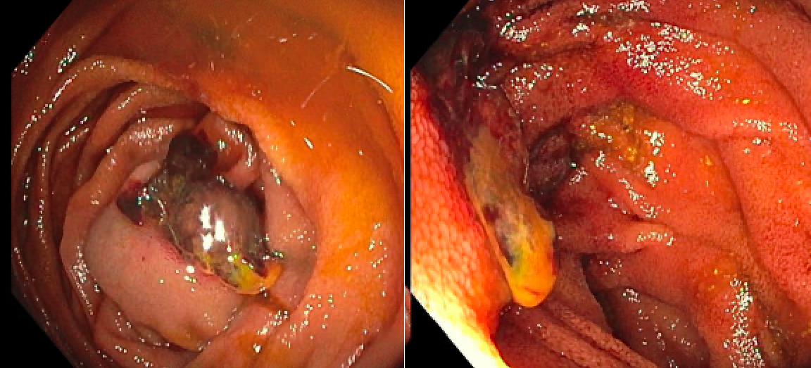
Methods: A 47-year-old man presented as a complex polytrauma with 33% total body surface area burns to his torso and extremities, requiring ICU stay, had melena and new transfusion requirements. An EGD demonstrated specs of hematin in the stomach and duodenum, without a source or other gross abnormalities. Colonoscopy revealed a rectal ulcer from a Flexiseal, but remainder of the colon and TI were normal. Due to ongoing anemia with overt bleeding, push enteroscopy was done which demonstrated a 1.5 cm raised, actively bleeding ulcerated lesion in the proximal jejunum but lesion was not biopsied due to bleeding. Exploratory laparotomy with small bowel resection and J-tube placement demonstrated a lesion that was polypoid with extensive surface ulceration and hemorrhage. The base of the lesion demonstrated a disrupted atypical vascular proliferation extending into the submucosa and partially involving the muscularis propria. Pathology revealed an angiosarcoma measuring 1.0x0.9x0.8 cm, positive for CD31. There was no evidence of recurrence or metastasis on imaging and pleural fluid cytology. Discussion: In the case of primary angiosarcomas of the small intestine, they have an extremely high metastasis rate of minimum 56.5% with common sites including the lung, liver, spleen and large intestines. Our case is unique in that, although patient was noted to have pleural effusions, cytology was negative for malignant cells, where as it is noted in literature, albeit in only handful of cases that respiratory failure due to pulmonary metastasis is a common cause of death in these patients. CT chest, abdomen and pelvis also did not reveal any evidence of metastasis to the lung or any other organs. It is also important to note that high suspicion is required to diagnose these tumors, gold standard remaining histological and immunohistochemical analysis.
Figure: 1.5 cm raised ulcerated lesion seen in the proximal jejunum
Disclosures: Sunanda Ellina indicated no relevant financial relationships. Ariana Tagliaferri indicated no relevant financial relationships. Silpa Choday indicated no relevant financial relationships. Aalam Sohal indicated no relevant financial relationships. Indu Srinivasan indicated no relevant financial relationships.
Sunanda Ellina, MD, Ariana R. Tagliaferri, MD, Silpa Choday, MD, Aalam Sohal, MD, Indu Srinivasan, MD. P2004 - A Rare Encounter: Localized Angiosarcoma of Jejunum, ACG 2025 Annual Scientific Meeting Abstracts. Phoenix, AZ: American College of Gastroenterology.

 photo")