University of Arizona College of Medicine, Phoenix Phoenix, AZ
Bianca Arellano, MD1, Asha Kurup, DO2, Vanessa Eller, MD1, Moti Salloum, MD2, Timothy Kuzel, MD2 1University of Arizona College of Medicine, Phoenix, Phoenix, AZ; 2University of Arizona College of Medicine, Phoenix VA Medical Center, Phoenix, AZ Introduction: Well-differentiated neuroendocrine tumors (NETs), previously known as carcinoid tumors, are rare with an approximate incidence of 4.7 per 100,000. The majority of NETs arise from the intestines or the lungs. Poorly differentiated neuroendocrine carcinoma (NEC) is an important distinction of pathology when classifying neuroendocrine neoplasms given their worse prognosis. The purpose of this case is to highlight the variability in histologic presentation, as well as propose an investigation for the occult primary site with known metastases to the liver and bone. Additionally, this case demonstrated a familial association with a first degree relative who died of carcinoid syndrome, without evidence of more common genetic mutations.
Case Description/
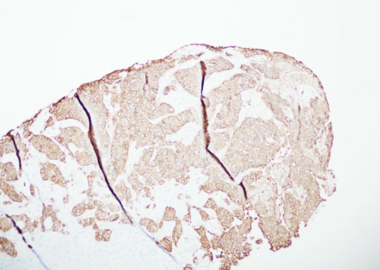
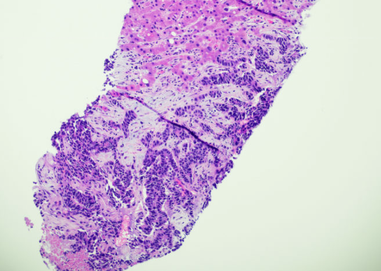
Methods: An 83 year old male presented to the hospital with acute hypoxic respiratory failure requiring oxygen. On arrival, patient was hypoxic, but other vital signs were stable. Admission labs were remarkable for ALP of 1248 U/L, AST 68 U/L, ALT of 52 U/L and a procalcitonin of 250 ng/ml. Chest imaging showed subcarinal adenopathy, pulmonary nodule, new osseous disease and multifocal liver metastasis. A thyroid ultrasound showed a new solid mass. The patient underwent echocardiogram which showed diastolic dysfunction andpulmonary hypertension Further workup included calcitonin of 5269 pg/ml, urine 5-HIAA of 140 mg/24 hours and chromagranin A of 192,900 ng/ml. The patient underwent liver biopsies resulting in pathology consistent with metastatic carcinoma with neuroendocrine features, including CDX-2 moderately positive and synaptophysin diffusely positive. The pathologist noted that the stain for Ki67 was nearly 100%, making well differentiated NET Grade 1, 2, 3 unlikely. Gallium Ga-68 dotatate scan performed showed uptake in the subcarinal node, liver and skeleton. Of note, no uptake was seen in the pancreas or bowel. Discussion: This case exemplifies an atypical pathology for neuroendocrine tumors and a potentially rare inheritance pattern of cancer. According to the pathology report, the tumor was originally identified as well-differentiated neuroendocrine tumor but later addended with large cell neuroendocrine carcinoma, noted to be very rare. The tumor appeared aggressive. Moreover, the patient unfortunately outlived his daughter. Limitations included the inability to obtain the daughter's medical records as well as the inability to obtain genetic testing on the patient. Potential genetic testing includes: MEN1, RET, VHL, NF1, TSC and IPMK.
Figure: H&E stain
Figure: Synaptophysin
Disclosures: Bianca Arellano indicated no relevant financial relationships. Asha Kurup indicated no relevant financial relationships. Vanessa Eller indicated no relevant financial relationships. Moti Salloum indicated no relevant financial relationships. Timothy Kuzel indicated no relevant financial relationships.
Bianca Arellano, MD1, Asha Kurup, DO2, Vanessa Eller, MD1, Moti Salloum, MD2, Timothy Kuzel, MD2. P2001 - A Unique Familial Presentation of Large Cell Neuroendocrine Carcinoma With Atypical Carcinoid Features, ACG 2025 Annual Scientific Meeting Abstracts. Phoenix, AZ: American College of Gastroenterology.