Frist College of Medicine at Belmont University Nashville, TN
Manav Patel, BS1, Ryan Roberts, MD2, William S. Barge, MD1 1Frist College of Medicine at Belmont University, Nashville, TN; 2Heritage Medical Associates, Nashville, TN Introduction: Leiomyosarcoma (LMS) is a rare and aggressive neoplasm of smooth-muscle origin that most often arises in the uterus or retroperitoneum. Colonic involvement is unusual and usually appears as a single symptomatic mass. Multifocal polypoid colonic lesions are uncommon and are only sparsely described. We report metastatic LMS of unknown primary origin detected during routine colonoscopy as three diminutive polyps that mimic tubular adenomas, illustrating the need to maintain a broad differential when widespread metastatic disease is suspected
Case Description/
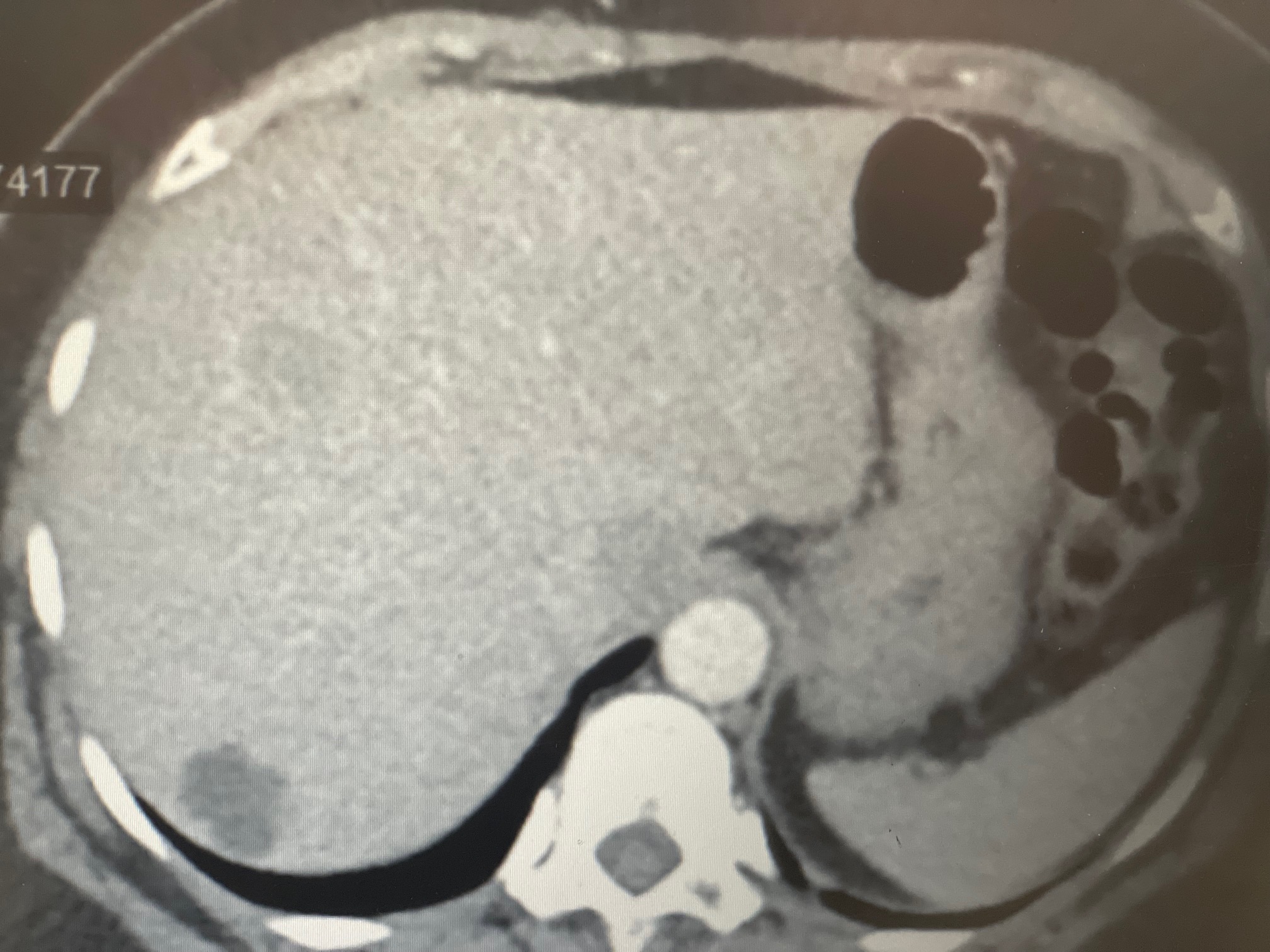
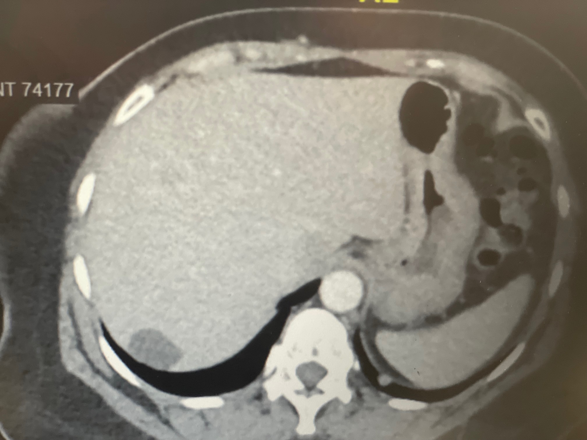
Methods: A fifty-nine-year-old woman presented with intermittent right-sided abdominal pain and uterine prolapse. Contrast-enhanced computed tomography revealed multiple hepatic lesions concerning for metastases, scattered pulmonary nodules, and cholelithiasis without cholecystitis; no primary tumor was seen. Colonoscopy identified three broad based sessile polyps measuring six millimeters each in the distal transverse colon, splenic flexure, and descending colon. All lesions appeared endoscopically similar to tubular adenomas and were removed with cold snare polypectomy. Histology showed pleomorphic spindle cells with abundant eosinophilic cytoplasm, marked nuclear atypia and immunohistochemistry was positive for desmin and smooth-muscle actin concerning for high grade metastatic LMS. Because of concurrent liver and lung disease the patient declined systemic therapy and chose palliative management. Written informed consent for publication was obtained. Discussion: Metastatic LMS rarely manifests as multiple diminutive colonic polyps that closely resemble benign adenomas. In this patient cross-sectional imaging suggested disseminated malignancy, yet the endoscopic appearance was misleadingly benign. Colonoscopy is often pursued in cases of metastatic disease of unknown primary, although typically to identify adenocarcinoma of the colon or a large colonic mass. Our case illustrates that even common colonoscopic findings may suggest a primary source of malignancy, even in the absence of mass lesion.
Figure: Cross-sectional imaging showing evidence of metastatic disease of unknown primary
Figure: Cross-sectional imaging showing evidence of metastatic disease of unknown primary
Disclosures: Manav Patel indicated no relevant financial relationships. Ryan Roberts indicated no relevant financial relationships. William Barge indicated no relevant financial relationships.
Manav Patel, BS1, Ryan Roberts, MD2, William S. Barge, MD1. P2551 - Metastatic Leiomyosarcoma Presenting as Three Benign-Appearing Colonic Polyps, ACG 2025 Annual Scientific Meeting Abstracts. Phoenix, AZ: American College of Gastroenterology.

.jpg "Manav Patel, BS photo")