University of Illinois College of Medicine Chicago, IL
Yousif Esho, MD1, Hetal Patel, MD1, Dexter E. Nwachukwu, DO1, James S. Love, MD1, Adam E. Mikolajczyk, MD2 1University of Illinois College of Medicine, Chicago, IL; 2University of Illinois, Chicago, IL Introduction: Appendiceal tumors are rare, accounting for 0.5–1% of intestinal neoplasms. Among these, neuroendocrine tumors (NETs) are the most common and are often incidentally found post-appendectomy. Due to vague symptoms, these tumors may go undiagnosed for years. This case highlights the importance of clinical vigilance, as early identification influences treatment, particularly decisions around hemicolectomy based on tumor size and invasion.
Case Description/
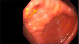
Methods: A 47-year-old female with a history of obesity and sleeve gastrectomy converted to Roux-en-Y gastric bypass presented for a screening colonoscopy. She had two prior unsuccessful colonoscopies due to poor bowel preparation. Before this third colonoscopy, she reported intermittent right lower quadrant pain. The colonoscopy showed mild mucosal edema and erythema near the appendiceal orifice without other significant findings (Figure 1) and biopsies revealed mild nonspecific chronic inflammation without dysplasia or malignancy.
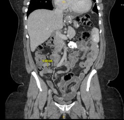
A dedicated abdominal CT scan and subsequent MRI revealed a thickened 1.4 cm appendiceal stump, which was unchanged from prior cross-sectional imaging 6 years earlier (Figure 2). With concern for a symptomatic appendiceal mucocele, she underwent elective laparoscopic appendectomy. Histopathology revealed a 2.6 cm well-differentiated appendiceal neuroendocrine tumor (NET), Ki-67 < 1%, grade 1, with submucosal and mesoappendiceal invasion. Surgical margins were negative for malignancy. Due to tumor size being greater than 2 cm and the presence of local invasion, oncology and surgical teams recommended a curative right hemicolectomy which was performed without complication.
Final pathology from the hemicolectomy revealed no residual tumor or lymph node involvement. Given the low-grade nature of the tumor and the absence of metastatic spread, the patient was managed conservatively with repeat surveillance imaging at regular intervals. She remains asymptomatic on follow-up with no evidence of recurrence.
Discussion: This case underscores the importance of carefully visualizing the appendiceal orifice during colonoscopy, especially in patients with prior poor preparation. It also highlights the need for comprehensive diagnostic workup, including cross-sectional imaging, when mucosal changes are seen at the orifice with symptoms. Although ulcerative colitis is most commonly associated with appendiceal orifice inflammation, appendiceal neuroendocrine tumors can rarely cause this finding when luminal obstruction occurs.
Figure: Figure 1 - Evidence of mild mucosal edema and erythema near the appendiceal orifice found during screening colonoscopy.
Figure: Figure 2 - CT imaging showing evidence of appendiceal stump thickening measuring 1.4 cm.
Disclosures: Yousif Esho indicated no relevant financial relationships. Hetal Patel indicated no relevant financial relationships. Dexter Nwachukwu indicated no relevant financial relationships. James Love indicated no relevant financial relationships. Adam Mikolajczyk indicated no relevant financial relationships.
Yousif Esho, MD1, Hetal Patel, MD1, Dexter E. Nwachukwu, DO1, James S. Love, MD1, Adam E. Mikolajczyk, MD2. P3032 - On the Outside, Looking In: An Uncommon Cause of Appendiceal Orifice Inflammation, ACG 2025 Annual Scientific Meeting Abstracts. Phoenix, AZ: American College of Gastroenterology.