Keck School of Medicine of the University of Southern California Los Angeles, CA
Mark C. Wang, MD1, Somaya Albhaisi, MBBCh, MPH1, Brandon B. Ge, MD1, Nikita Saeedi, BS1, Theodor Griggs, MD, PhD2 1Keck School of Medicine of the University of Southern California, Los Angeles, CA; 2Los Angeles General Medical Center, Los Angeles, CA Introduction: While varices are a common cause of brisk esophageal bleeding in patients with cirrhosis, other rarer sources should still be considered, especially in critically ill patients. Here, we describe a patient with cirrhosis who developed a brisk esophageal bleed from a left gastric artery pseudoaneurysm in the setting of severe esophagitis.
Case Description/
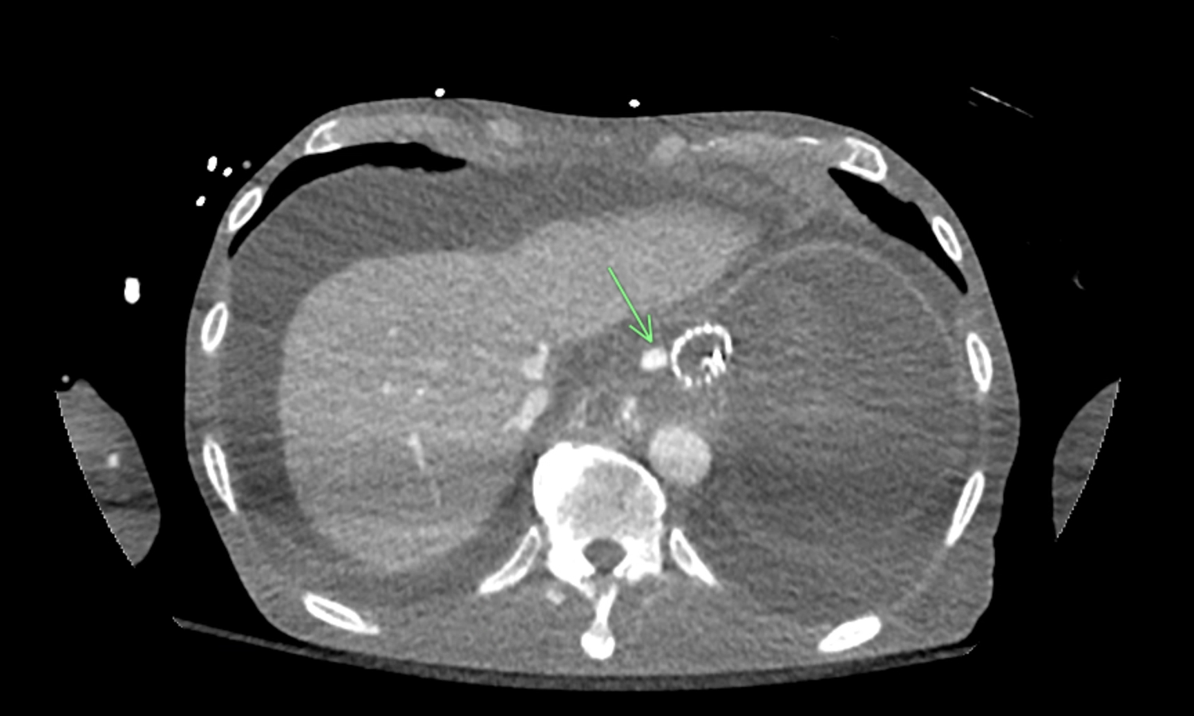
Methods: A 53-year-old male with chronic hepatitis B, cirrhosis, and amphetamine use disorder presented after being found unresponsive with melena and coffee ground emesis. He had a recent history of admissions for dark emesis thought to be secondary to amphetamine-induced esophagitis, though no previous endoscopies were performed. He was initially stable with labs significant for hemoglobin 4.9 g/dL, lactate 3.1, and severe coagulopathy. However, he then developed shock and hematemesis, necessitating ICU transfer. EGD revealed severe esophagitis in the distal esophagus. A source of brisk bleeding was seen in the distal esophagus and believed to be a varix. However, it was not amenable to ligation, so hemostatic powders were applied. CT angiography showed engorged varices without evidence of arterial hemorrhage. The patient initially improved but began to have hematemesis again one day later. A second EGD was performed, again finding active hemorrhage in the distal esophagus surrounded by worsening esophagitis. Several interventions, including hemostasis powders, epinephrine, and ethanolamine, were unsuccessful, so a fully-covered stent was placed to tamponade the bleed. Subsequent decompensation prompted a second CT angiography (Figure 1), revealing active extravasation from a pseudoaneurysm in the gastroesophageal branch of the left gastric artery, which was subsequently embolized. The patient improved and remained stable for three days; however, subsequent decompensation led to a transition to comfort care. Discussion: In this case, a left gastric artery pseudoaneurysm caused a brisk esophageal bleed that was originally suspected to be variceal after initial EGD and imaging. Left gastric artery pseudoaneurysms are a rare cause of upper GI bleed and usually form due to trauma, pancreatitis, and atherosclerosis. It is possible that the pseudoaneurysm developed due to severe local inflammation from the amphetamine-induced esophagitis. Worsening esophagitis and coagulopathy likely caused the pseudoaneurysm to rupture, resulting in a bleed refractory to endoscopic intervention and requiring embolization.
Figure: Figure 1: CT Angiography Demonstrating the Pseudoaneurysm in the Gastroesophageal Branch of the Left Gastric Artery
Disclosures: Mark Wang indicated no relevant financial relationships. Somaya Albhaisi indicated no relevant financial relationships. Brandon Ge indicated no relevant financial relationships. Nikita Saeedi indicated no relevant financial relationships. Theodor Griggs indicated no relevant financial relationships.
Mark C. Wang, MD1, Somaya Albhaisi, MBBCh, MPH1, Brandon B. Ge, MD1, Nikita Saeedi, BS1, Theodor Griggs, MD, PhD2. P3116 - Beyond Varices: Left Gastric Pseudoaneurysm as a Rare Cause of Upper GI Bleed in Cirrhosis, ACG 2025 Annual Scientific Meeting Abstracts. Phoenix, AZ: American College of Gastroenterology.

 photo")