Corewell Health William Beaumont University Hospital Royal Oak, MI
Ahmed Aref, MD1, Nathanial Bartosek, MD2, Mujtaba Moazzam, MD1, Fady Banno, MD1, Nishant Aggarwal, MD1, Laith H. Jamil, MD, FACG3 1Corewell Health William Beaumont University Hospital, Royal Oak, MI; 2Corewell Health East William Beaumont University Hospital, Royal Oak, MI; 3Oakland University William Beaumont School of Medicine, Rochester, MI Introduction: Cytomegalovirus (CMV) colitis typically affects immunocompromised patients, although it’s a rare cause of gastrointestinal (GI) bleeding in immunocompetent patients. CMV rarely presents as a mass. We report a case of CMV infection in a critically ill immunocompetent patient presenting as a mass-like lesion at the ileocecal valve.
Case Description/
Methods: A 47-year-old male with no known history of immunodeficiency presented with respiratory failure and acute respiratory distress syndrome (ARDS). Extensive workup was inconclusive, but the presentation raised concern for cryptogenic organizing pneumonia or acute interstitial pneumonitis. He required intubation and mechanical ventilation and was subsequently placed on veno-venous extracorporeal membrane oxygenation.
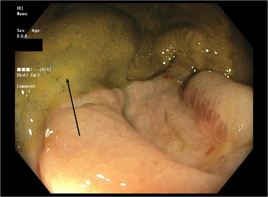
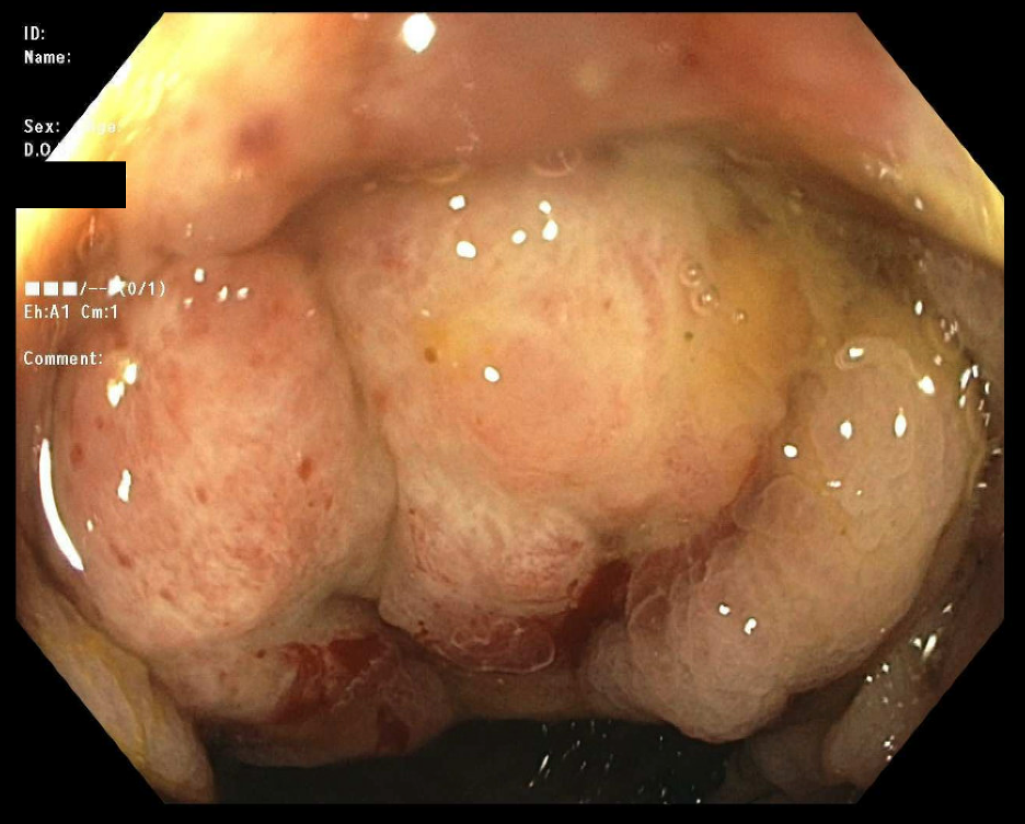
He developed hematochezia with a hemoglobin from drop from 10g/dL to 8.3 g/dL. Esophagogastroduodenoscopy was unremarkable. Colonoscopy revealed a 3 × 3.5 cm ulcer in the hepatic flexure, which was the likely source of bleeding (Figure 1), and a friable, multilobular mass at the ileocecal valve extending into the ascending colon which was biopsied (Figure 2). Mesenteric angiography revealed active extravasation at the hepatic flexure, site of the prior ulcer, with embolization of two vasa recta branches, after which his hematochezia resolved.
Histopathology demonstrated ulcerated small intestinal mucosa, with immunohistochemistry positive for CMV. The initial serum CMV viral load was 6,910 IU/mL that decreased after treatment with ganciclovir to 265 IU/mL. A month later, mesenteric angiography for recurrent hematochezia showed ileocecal bleeding, with embolization of 3 bleeding vessels. His respiratory status did not improve over a hospital course of about 3 months and his family elected to pursue comfort care. He subsequently passed away. Discussion: Although CMV primarily affects immunocompromised individuals, many immunocompetent patients who present with CMV colitis have some degree of immunosuppression, such as critical illness in our patient. GI CMV infections manifest as ulcerative lesions, but mass lesions have also been reported. Commonest sites are right colon and ileocecal valve. Studies have reported mortality rates as high as 71.4% in critically ill, immunocompetent patients with CMV colitis, even with ganciclovir This case highlights the need to consider CMV in critically ill patients with GI bleeding and mass lesions, even without known immunodeficiency.
Figure: Figure 1: Ulcer at the hepatic flexure
Figure: Figure 2: Ileocecal mass
Disclosures: Ahmed Aref indicated no relevant financial relationships. Nathanial Bartosek indicated no relevant financial relationships. Mujtaba Moazzam indicated no relevant financial relationships. Fady Banno indicated no relevant financial relationships. Nishant Aggarwal indicated no relevant financial relationships. Laith Jamil indicated no relevant financial relationships.
Ahmed Aref, MD1, Nathanial Bartosek, MD2, Mujtaba Moazzam, MD1, Fady Banno, MD1, Nishant Aggarwal, MD1, Laith H. Jamil, MD, FACG3. P3479 - Cytomegalovirus Enterocolitis Presenting as a Mass-Like Lesion at the Ileocecal Valve in a Critically Ill Immunocompetent Patient, ACG 2025 Annual Scientific Meeting Abstracts. Phoenix, AZ: American College of Gastroenterology.