University of Miami Miller School of Medicine at JFK Medical Center Atlantis, FL
Bebika Subedi, MD, Kailee Vickaryous, DO, Dipen Dhakal, MD University of Miami Miller School of Medicine at JFK Medical Center, Atlantis, FL Introduction: Trichuriasis, caused by the whipworm Trichuris trichiura, is an intestinal infection acquired via ingestion of embryonated eggs from contaminated environments. Though rare in developed countries (~100 cases per 100,000 in the U.S.), it remains common in tropical and subtropical regions. Clinical presentations vary from asymptomatic to severe cases involving diarrhea, anemia, and malnutrition. The sensitivity of stool examinations, such as the Kato-Katz technique, varies widely (42.8% to 95%), and false negatives are common, particularly in low grade infections or intermittent egg shedding. When stool studies are inconclusive, colonoscopy can offer direct visualization of adult worms and enables therapeutic interventions like worm removal. This case highlights colonoscopy’s role in diagnosing T. trichiura in a patient with malnutrition and severe anemia.
Case Description/
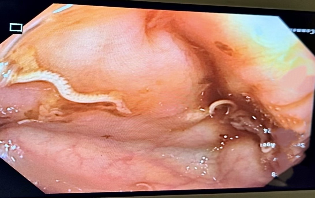
Methods: A 29 year old male with history of chronic alcohol use presented with two months of malaise, dizziness, unintentional weight loss, poor appetite, and dark stools. He denied abdominal pain or any other systemic symptoms. Labs showed severe microcytic anemia (Hb 3.3 g/dL, MCV 64.1 fL), requiring transfusion and further workup. Stool ova and parasite tests were initially negative. Colonoscopy revealed live whipworms embedded in the cecal mucosa around 4 cm in length, which was later confirmed by pathology. EGD was unremarkable except for a small hiatal hernia. Imaging showed fatty liver, consistent with alcohol use. He was treated with Albendazole 400 mg for 3 days, with notable symptom improvement and hemoglobin stabilization to 8 g/dL. He was discharged in stable condition with outpatient follow-up for eradication confirmation. Discussion: Although uncommon in developed nations, T. trichiura can lead to significant anemia and nutritional deficiency when parasite burden is high. Stool examinations, while commonly used, may yield false negatives, necessitating additional diagnostic approaches. Colonoscopy offers a valuable alternative by enabling direct detection and simultaneous treatment. Its role is particularly critical in diagnosing parasitic infections when standard stool analyses fail, as demonstrated in this case. In patients with negative stool studies but high clinical suspicion, colonoscopy is critical for timely diagnosis and effective management.
Figure: Colonoscopy showing a live adult Trichuris trichiura attached to the cecal mucosa.
Disclosures: Bebika Subedi indicated no relevant financial relationships. Kailee Vickaryous indicated no relevant financial relationships. Dipen Dhakal indicated no relevant financial relationships.
Bebika Subedi, MD, Kailee Vickaryous, DO, Dipen Dhakal, MD. P3477 - The Hidden Worms: Colonoscopy as a Key Diagnostic Tool for Parasitic Infestation, ACG 2025 Annual Scientific Meeting Abstracts. Phoenix, AZ: American College of Gastroenterology.