St. Joseph's University Medical Center Paterson, NJ
Lefika Bathobakae, MD, MPH1, Eliesther Rivera, MD2, Rammy Bashir, MD, MSc3, Reshma John, MD, MPH1, Irhoboudu Atogwe, MD4, Derya Mücahit, DO5, Kamal Amer, MD6, Walid Baddoura, MD1 1St. Joseph's University Medical Center, Paterson, NJ; 2American University of Antigua College of Medicine, Paterson, NJ; 3Norwalk Hospital/Yale University, Norwalk, CT; 4Albert Einstein Medical Center, Paterson, NJ; 5Newark Beth Israel Medical Center, Paterson, NJ; 6St. Joseph’s University Medical Center, Paterson, NJ Introduction: Duodenal pseudomelanosis is an extremely rare, benign condition typified by the presence of dark pigmented spots in the duodenal mucosa.1,2 First described in 1976, pseudomelanosis duodeni continues to be an incidental finding on endoscopy and has an unclear clinical significance.3,4Herein, we describe a case series of duodenal pseudomelanosis diagnosed at our institution. It is important for clinicians to recognize duodenal pseudomelanosis, given its tendency to mimic serious conditions, such as melanoma, metastatic disease, and drug-induced pigmentation.
Case Description/
Methods: Case 1
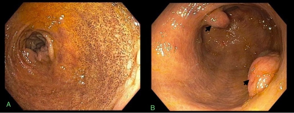
A 74-year-old man with a history of ESRD on HD, type 2 diabetes, and HTN was brought to the ED for acute-onset epigastric pain and melena. Triage labs: hemoglobin, 5.3 g/dL, and BUN, 90 mg/dL. EGD revealed a small hiatal hernia, three gastric nodules with no stigmata of recent bleeding, and distal antral and duodenal melanosis (Figure 1). Duodenal bulb specimens showed black granular pigment within the macrophages of the lamina propria, which stained positive for iron, consistent with pseudomelanosis.
Case 2
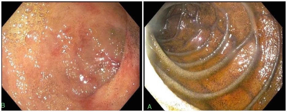
An 86-year-old man with a history of ESRD on HD, HTN, and obscure GI bleeding was brought to the ED for altered mental status and fever. Triage blood tests: low hemoglobin (4.7 g/dL) and leukocytosis (WBC, 14.4 x 10³/mm3). Prior endoscopic procedures were non-revealing. EGD showed erythematous duodenopathy and severe melanosis in the second portion of the duodenum (Figure 2). No biopsies were obtained for histopathological analysis and the patient was managed conservatively.
Case 3
A 79-year-old woman with a history of acute ischemic stroke, HTN, type 2 diabetes, and IDA requiring iron sucrose infusions presented to the ED complaining of acute-onset abdominal pain. Labs: low hemoglobin, 7.8 g/dL. Imaging revealed prominent colitis and proctitis without perforation or abscess formation. EGD showed a small hiatal hernia, atrophic bile gastritis, and diffuse duodenal melanosis. Histopathology findings were consistent with duodenal pseudomelanosis. Discussion: The etiopathogenesis of duodenal pseudomelanosis is not fully understood, but it has been associated with some chronic illnesses and certain medications. The diagnosis of pseudomelanosis is made based on endoscopic findings of dark-speckled mucosal pigmentation and often confirmed on histology. Recognizing this novel entity is crucial for accurate diagnosis, appropriate management of associated conditions, and avoiding unnecessary interventions.
Figure: Figure 1. Endoscopic image showing multiple dark brown spots in the duodenal bulb (panel A) and gastric nodules (black arrows).
Figure: Figure 2. Endoscopic images showing severe melanosis of the second portion of the duodenum (panel A) and diffuse moderately erythematous mucosa of the duodenal bulb (panel B). No stigmata of recent bleeding were found in the duodenal bulb.
Disclosures: Lefika Bathobakae indicated no relevant financial relationships. Eliesther Rivera indicated no relevant financial relationships. Rammy Bashir indicated no relevant financial relationships. Reshma John indicated no relevant financial relationships. Irhoboudu Atogwe indicated no relevant financial relationships. Derya Mücahit indicated no relevant financial relationships. Kamal Amer indicated no relevant financial relationships. Walid Baddoura indicated no relevant financial relationships.
Lefika Bathobakae, MD, MPH1, Eliesther Rivera, MD2, Rammy Bashir, MD, MSc3, Reshma John, MD, MPH1, Irhoboudu Atogwe, MD4, Derya Mücahit, DO5, Kamal Amer, MD6, Walid Baddoura, MD1. P4096 - Pigment on the Path: A Case Series of Duodenal Pseudomelanosis From a Single Institution, ACG 2025 Annual Scientific Meeting Abstracts. Phoenix, AZ: American College of Gastroenterology.