Penn State Health Milton S. Hershey Medical Center Hershey, PA
Anil Bhatnagar, MD1, Matthew O'Neil, MD1, Justin Canakis, DO1, Jennifer Maranki, MD2 1Penn State Health Milton S. Hershey Medical Center, Hershey, PA; 2Penn State College of Medicine, Hershey, PA Introduction: Superior mesenteric artery syndrome (SMA) syndrome is an uncommon complication of spinal deformity corrective surgery with an estimated incidence of 2% following scoliosis surgery in adult patients. It is thought to be the result of the aortomesenteric angle narrowing due to the lengthening of the spine and loss of retroperitoneal fat, leading to extrinsic compression of the duodenum. We present a case of SMA syndrome secondary to scoliosis repair surgery to highlight this patient’s unique imaging findings, and to emphasize the importance of considering SMA syndrome in the differential for postoperative gastrointestinal complaints.
Case Description/
Methods: A 21-year-old male developed superior mesenteric artery (SMA) syndrome as a complication of posterior spinal fusion performed for progressive thoracogenic scoliosis. He had a history of thoracotomy and repair of severe aortic coarctation during infancy, which led to progressive spinal deformity over time. By early adulthood, he developed a 65-degree thoracic curvature, prompting posterior T12-L1 spinal fusion.
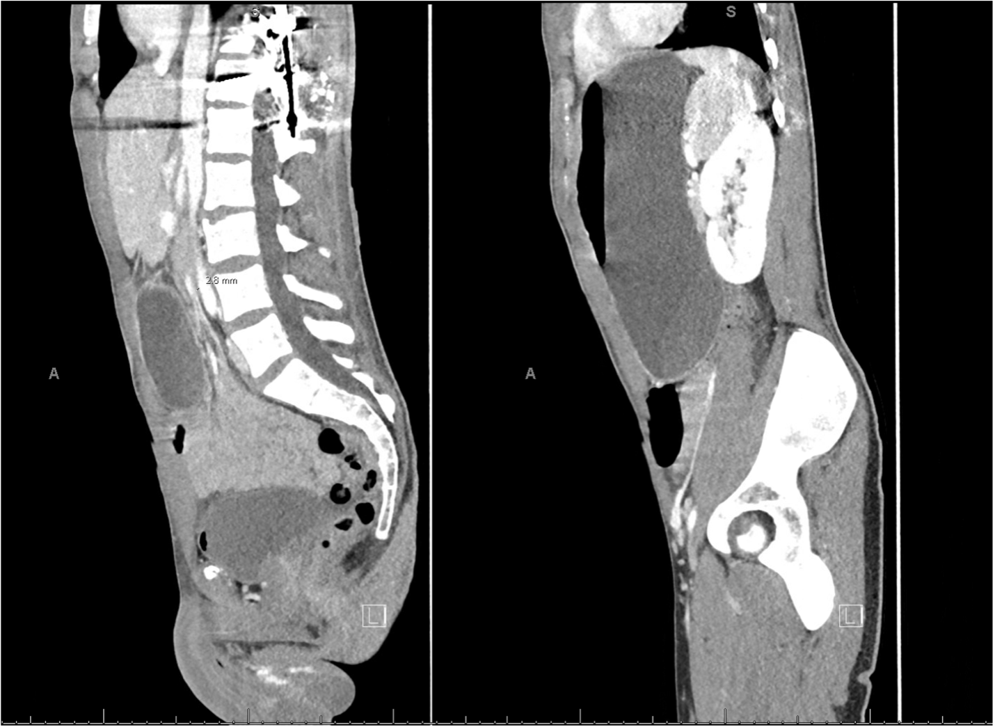
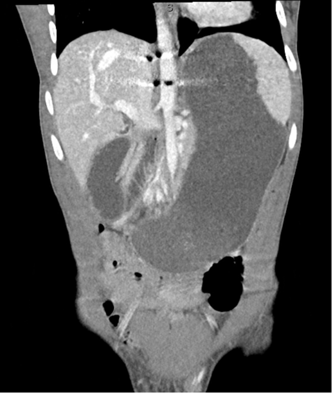
He was discharged uneventfully on postoperative day three. However, on postoperative day six, he re-presented complaining of abdominal pain, non-bloody emesis, and “feeling like something [was] stuck in [his] belly.” CT abdomen and pelvis with contrast demonstrated marked gastric distension and abrupt narrowing of the third portion of the duodenum with decompressed bowel distally – findings suspicious for SMA syndrome (Figure 1, Figure 2).
The patient was initially managed conservatively with nasogastric decompression and nasojejunal feeding. However, due to persistent symptoms, he ultimately underwent laparoscopic intestinal derotation and gastrojejunostomy (GJ) tube placement. He was discharged with a combination of tube feeds and oral supplementation. Discussion: In this rare case, this patient ultimately underwent intestinal derotation and gastrojejunostomy tube placement after conservative therapy had failed. After a few months, his GJ tube was removed and he reports his gastrointestinal symptoms have improved to his baseline prior to scoliosis surgery. This case highlights SMA syndrome as a rare but important postoperative complication of scoliosis corrective surgery. Presenting symptoms may mimic ileus or other nonspecific postoperative gastrointestinal complaints but should prompt early cross-sectional imaging when persistent.
Figure: Figure 1. Sagittal view demonstrating severe narrowing of the aortomesenteric angle (left) and marked gastric distension due to superior mesenteric artery (SMA) syndrome (right).
Figure: Figure 2. Coronal view demonstrating marked gastric distension and narrowing of the D3 duodenal segment due to SMA syndrome.
Disclosures: Anil Bhatnagar indicated no relevant financial relationships. Matthew O'Neil indicated no relevant financial relationships. Justin Canakis indicated no relevant financial relationships. Jennifer Maranki indicated no relevant financial relationships.
Anil Bhatnagar, MD1, Matthew O'Neil, MD1, Justin Canakis, DO1, Jennifer Maranki, MD2. P4161 - Distinctive Imaging of Superior Mesenteric Artery Syndrome Following Scoliosis Repair, ACG 2025 Annual Scientific Meeting Abstracts. Phoenix, AZ: American College of Gastroenterology.