Geisinger Commonwealth School of Medicine Scranton, PA
Hannah Anderson, MS1, Christopher D.. Manko, BS1, Michelle Bernshteyn, MD2, Subash Ghimire, MD2, Ashit B.. Sarker, MD, PhD3, Joyson Poulose, MD4, Michael Georgetson, MD, FACG2 1Geisinger Commonwealth School of Medicine, Scranton, PA; 2Guthrie Robert Packer Hospital, Department of Gastroenterology, Sayre, PA; 3Guthrie Robert Packer Hospital, Department of Pathology, Sayre, PA; 4Guthrie Robert Packer Hospital, Department of Hematology/Oncology, Sayre, PA Introduction: Gastric cancer is the fourth most common cause of cancer death for both men and women (1). Leiomyosarcoma (LMS) is a malignant smooth muscle tumor that very rarely affects the stomach. We present here a case of a male patient with LMS of the stomach. This case highlights the importance of follow-up care, as this diagnosis was made only on follow-up endoscopy (EGD).
Case Description/
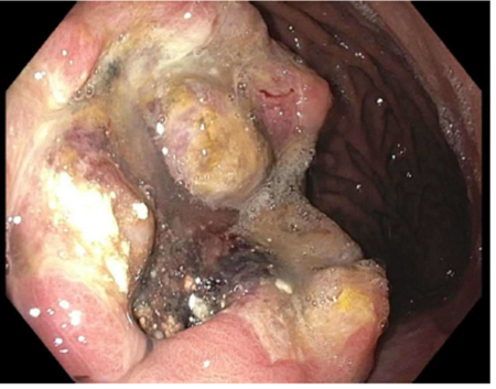
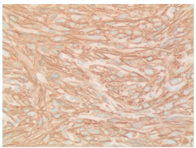
Methods: The patient is a 58-year-old male who presented to the emergency department for a sensation of rapid heartbeat, dark tarry stools for a month, and postprandial epigastric pain radiating to the back. Workup was positive for anemia and atrial fibrillation with rapid ventricular response. Upper EGD revealed a gastric body ulcer with predominantly necroinflammatory debris, negative for H. pylori and intestinal metaplasia. Repeat EGD in 12 weeks was planned. Two months later, the patient noted fatigue, outpatient CBC again revealed anemia, and the patient was readmitted. Repeat EGD showed a non-obstructing gastric ulcer with biopsy exhibiting spindle cells diffusely positive for smooth muscle actin. Unfortunately, diagnosis of high-grade gastric LMS was made and abdominal CT showed a mass along the gastric fundus, involvement of a periportal lymph node and multiple hepatic lesions. Initial treatment of doxorubicin and trabectedin was pursued, with the patient later opting for home hospice care. Discussion: This report describes a rare case of gastric LMS presenting as a gastric ulcer. LMS most often presents in the retroperitoneum or uterus (2) and, to the best of our knowledge, this is the first described case of metastatic gastric LMS presenting as an ulcer, without a mass or polypoid lesion visible on EGD. Moreover, while H. pylori has been suspected to play a role in the development of gastric LMS (3), our patient was H. pylori negative. Initial biopsy of the gastric ulcer was negative for neoplasm and only follow-up EGD during inpatient management caught the diagnosis of LMS. This point emphasizes the importance of providers following up with gastroenterology concerns, as false negative results are possible, even with biopsy.
References: 1. Ilic M, Ilic I. Epidemiology of stomach cancer. World J Gastroenterol. 2022 Mar 28;28(12):1187–203. 2. Menon G, Mangla A, Yadav U. Leiomyosarcoma. In: StatPearls. Treasure Island (FL): StatPearls Publishing; 2025 Jan-. 3. Wang T, Zreik R, Leng B. Primary Gastric Leiomyosarcoma: A Rare Case. Cureus. 2023 Nov 27;15(11):e49510.
Figure: Figure 1. Endoscopy showing gastric body ulcer
Figure: Figure 2. Histology slide of gastric ulcer biopsy with positive smooth muscle actin stain
Disclosures: Hannah Anderson indicated no relevant financial relationships. Christopher Manko indicated no relevant financial relationships. Michelle Bernshteyn indicated no relevant financial relationships. Subash Ghimire indicated no relevant financial relationships. Ashit Sarker indicated no relevant financial relationships. Joyson Poulose indicated no relevant financial relationships. Michael Georgetson indicated no relevant financial relationships.
Hannah Anderson, MS1, Christopher D.. Manko, BS1, Michelle Bernshteyn, MD2, Subash Ghimire, MD2, Ashit B.. Sarker, MD, PhD3, Joyson Poulose, MD4, Michael Georgetson, MD, FACG2. P4276 - A Rare Case of Gastric Leiomyosarcoma in a Male Patient, ACG 2025 Annual Scientific Meeting Abstracts. Phoenix, AZ: American College of Gastroenterology.