University of Central Florida, HCA Healthcare GME Pensacola, FL
Priya Kumari Maheshwari, MD1, Parth Desai, DO2, Jillian R. Grau, MD3, Hiral Shah, MD4 1University of Central Florida, HCA Healthcare GME, Pensacola, FL; 2Lehigh Valley Health Network, Allentown, PA; 3HNL Lab Medicine, Allentown, PA; 4Lehigh Valley Health Network/Jefferson Health, Center Valley, PA Introduction: Calcifying fibrous tumor (CFT) is a rare, benign mesenchymal neoplasm most commonly found in the pleura, soft tissues, or peritoneum. Gastrointestinal involvement is rare, especially in the stomach. These lesions are typically asymptomatic and often discovered incidentally. On imaging and endoscopy, they may mimic other subepithelial gastric lesions such as gastrointestinal stromal tumors (GISTs), leiomyomas, or duplication cysts. We present a case of a gastric CFT incidentally identified during evaluation for gastroesophageal reflux disease (GERD), which was successfully treated with endoscopic submucosal dissection (ESD).
Case Description/
Methods: A 52-year-old woman with a history of hypothyroidism, papillary thyroid carcinoma post thyroidectomy, and GERD presented with persistent reflux symptoms. An esophagogastroduodenoscopy (EGD) revealed an incidental 2.5 cm subepithelial lesion (SEL) along the lesser curvature of the stomach near the angularis, with a smooth, non-ulcerated surface. Endoscopic ultrasound (EUS) demonstrated a hypoechoic lesion with a calcified rim and cystic center, confined to the second layer with an intact muscularis propria. Differential considerations included a duplication cyst, leiomyoma, GIST, or calcified lipomatous lesion.
Due to limitations in visualization from acoustic shadowing and potential infection risk, fine-needle aspiration was deferred. Serial imaging over 15 months showed a stable lesion. After multidisciplinary discussions, the patient elected to undergo ESD for diagnostic and therapeutic purposes. A technically complex ESD was performed, achieving en bloc resection of a 5 cm lesion. Histopathology confirmed a benign calcifying fibrous tumor with negative peripheral margins and focal abutment of the deep margin. The patient tolerated the procedure well and was discharged with plans for close follow-up. Discussion: Although rare, gastric CFTs present a unique diagnostic challenge due to their nonspecific imaging characteristics and resemblance to more aggressive lesions. Definitive preoperative diagnosis is not possible without tissue resection. While CFTs have no known malignant potential, accurate identification is essential to avoid unnecessary surgical intervention. Complete excision is considered curative, and recurrence is rare. This case emphasizes the importance of considering CFT in the differential diagnosis of gastric subepithelial lesions and illustrates the utility of advanced endoscopic techniques in managing rare benign gastric tumors.
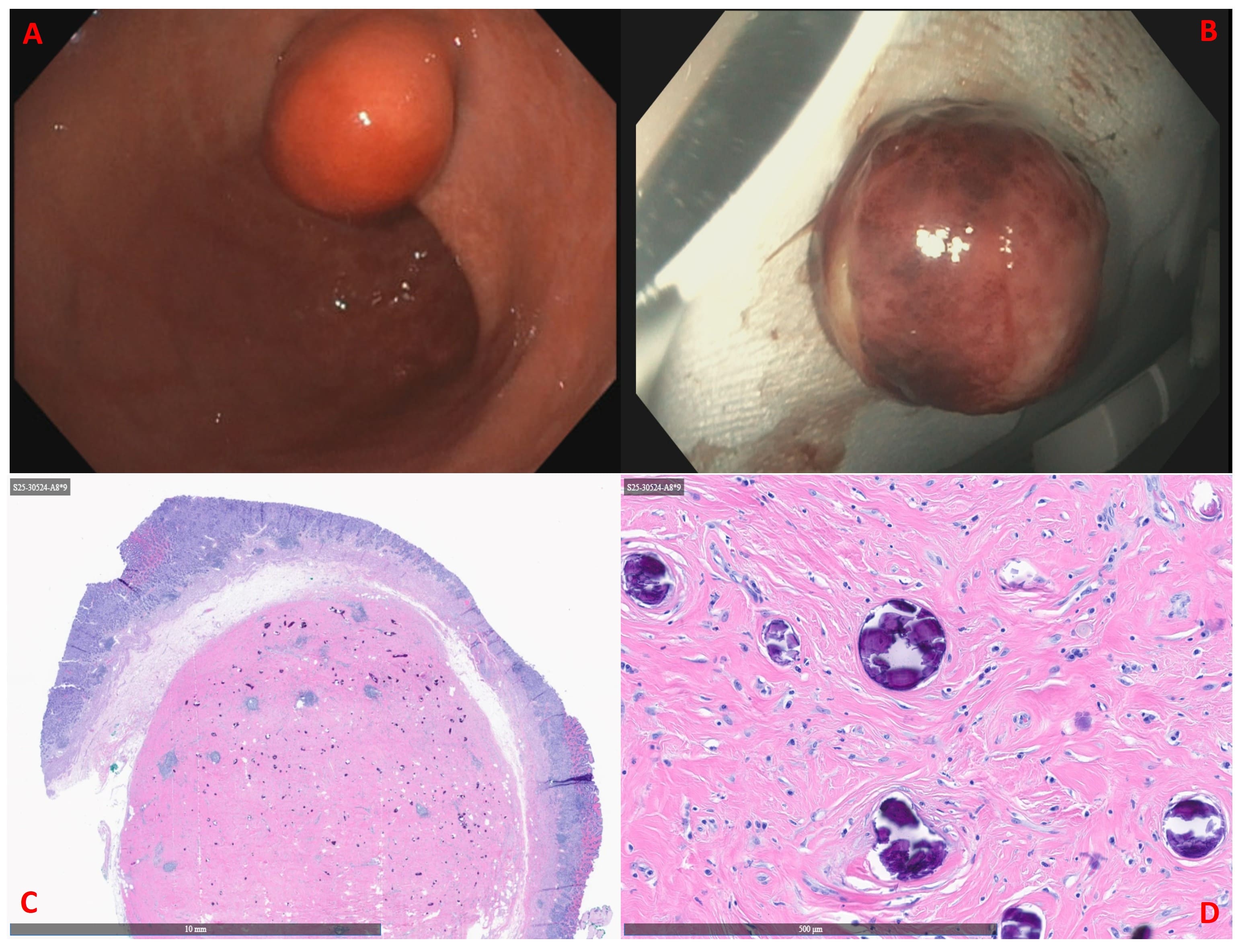
Figure: Figure 1A: Endoscopic images showing a well-circumscribed, smooth, subepithelial lesion located along the lesser curvature of the stomach. Figure 1B: Gross view of the lesion following en bloc resection by endoscopic submucosal dissection. Figure 1C-D: H&E stain showing dense hyalinized stroma with psammomatous calcifications and scattered spindle cells, consistent with calcifying fibrous tumor.
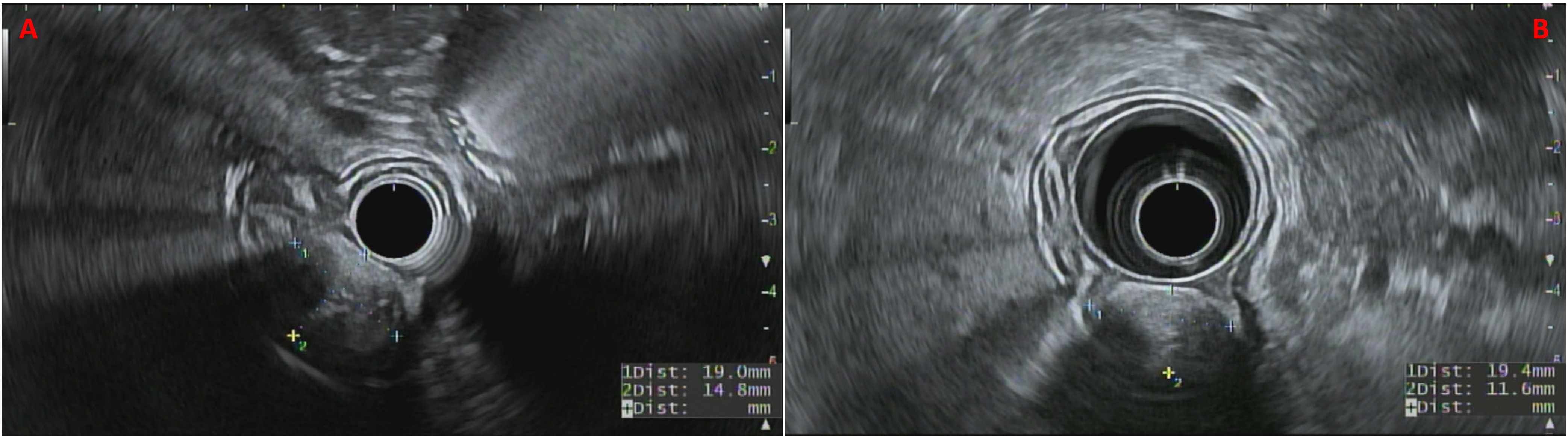
Figure: Figure 2A-B: Endoscopic ultrasound (EUS) images showing a well-circumscribed, hypoechoic lesion arising from the second layer (deep mucosa), with an intact muscularis propria.
Disclosures: Priya Kumari Maheshwari indicated no relevant financial relationships. Parth Desai indicated no relevant financial relationships. Jillian Grau indicated no relevant financial relationships. Hiral Shah: Abbvie – Speakers Bureau.
Priya Kumari Maheshwari, MD1, Parth Desai, DO2, Jillian R. Grau, MD3, Hiral Shah, MD4. P4211 - Gastric Calcifying Fibrous Tumor: An Incidental Subepithelial Lesion Managed by Endoscopic Submucosal Dissection, ACG 2025 Annual Scientific Meeting Abstracts. Phoenix, AZ: American College of Gastroenterology.

 photo")