University of Kansas School of Medicine - Wichita Wichita, KS
John Thesing, DO1, Meagan H. Phox, DO2, Audrey Lazenby, MD3, Aaron Brown, MD4, John Thesing, MD5 1University of Kansas School of Medicine - Wichita, Wichita, KS; 2University of Kansas School of Medicine, Wichita, KS; 3University of Nebraska Medical Center, Omaha, NE; 4Holton Community Hospital, Holton, KS; 5Holton Community Hospital, Leawood, KS Introduction: Ménétrier disease is a rare hypertrophic gastropathy marked by enlarged gastric folds and protein loss. It typically affects men aged 30–60 but can occur in children, where it is often self-limiting. It is associated with gastric cancer and may follow infections like Helicobacter pylori (H. pylori) or cytomegalovirus (CMV). We present a case of a male in his 20s who presented with an acute Ménétrier-like condition.
Case Description/
Methods: A 27-year-old male with no prior medical history presented with acute vomiting and abdominal pain. Initial labs showed elevated hemoglobin (Hgb) of 19.3 g/dL and white blood cell count (WBC) of 15.3 x10³/μL; computed tomography (CT) abdomen/pelvis was unremarkable. He was treated with IV fluids and antiemetics and discharged.
Nine days later, he returned with worsening symptoms and new-onset hand and pedal edema. Labs again showed elevated Hgb (18.3 g/dL), WBC (17.9 x10³/μL), and hypoalbuminemia (1.5 g/dL, previously 3.8 g/dL). Urinalysis was negative for proteinuria. Inflammatory markers were normal. Repeat CT showed circumferential gastric wall thickening, IV contrast enhancement of the gastric wall, ascites, and soft tissue edema. He denied any recent changes in medications or supplements. He was admitted and underwent esophagogastroduodenoscopy (EGD).
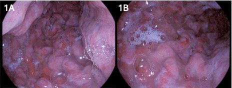
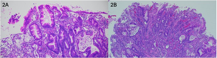
EGD revealed thickened gastric folds with mucosal sloughing, hyperemia, and thick mucus (Figure 1). Full thickness biopsies using a cold snare were obtained. Biopsies from the body and fundus showed foveolar hyperplasia and active chronic gastritis with erosions (Figure 2). Small bowel biopsies were unremarkable. Stains for H. pylori, CMV, Herpes Simplex Virus, Epstein Barr virus, and adenovirus were negative. Stool PCR testing for enteric pathogens was also negative.
He was managed symptomatically with scopolamine, sucralfate, PPI, and a high-protein diet. His albumin rose to 3.0 g/dL by week 3 post-discharge and normalized to 3.7 g/dL by week 7. Discussion: This case shows an acute, atypical presentation of Ménétrier-like disease in a young adult, resembling pediatric cases but without an obvious, infectious trigger. Though rare, it should be considered in patients with hypoalbuminemia and gastric hypertrophy. When suspected, full-thickness biopsies with a snare are preferred over forceps to assess mucosal architecture. Continued surveillance is recommended due to the risk of recurrence or progression.
Figure: Figure 1: Endoscopic view demonstrating circumferential hypertrophic rugae and hyperemic mucosa without ulceration or bleeding with abundant, thick mucus.
Figure: Figure 2: Marked foveolar hyperplasia with elongation, tortuosity, and loss of mucin in the foveolae with active, chronic inflammation.
Disclosures: John Thesing indicated no relevant financial relationships. Meagan Phox indicated no relevant financial relationships. Audrey Lazenby indicated no relevant financial relationships. Aaron Brown indicated no relevant financial relationships. John Thesing indicated no relevant financial relationships.
John Thesing, DO1, Meagan H. Phox, DO2, Audrey Lazenby, MD3, Aaron Brown, MD4, John Thesing, MD5. P4210 - Giant Gastric Folds, Subtle Signs: Ménétrier-Like Disease in a Young Adult, ACG 2025 Annual Scientific Meeting Abstracts. Phoenix, AZ: American College of Gastroenterology.