University of Texas Health San Antonio San Antonio, TX
Parth Patel, MD, MBA1, Saatchi Kuwelker, MD1, Sherif Ibrahim, MD2, Hari Sayana, MD1 1University of Texas Health San Antonio, San Antonio, TX; 2University of South Carolina School of Medicine, San Antonio, TX Introduction: Linitis plastica is a phenotype of gastric carcinoma that occurs in 7 to 10% of all primary gastric cancers. This malignant lesion is typically asymptomatic until advanced stages due to the reduced sensitivity of space occupying lesions within the stomach. Once symptomatic, patients present with dyspepsia, vomiting, dysphagia, and unexpected weight loss. We present a case of rapidly progressing linitis plastica.
Case Description/
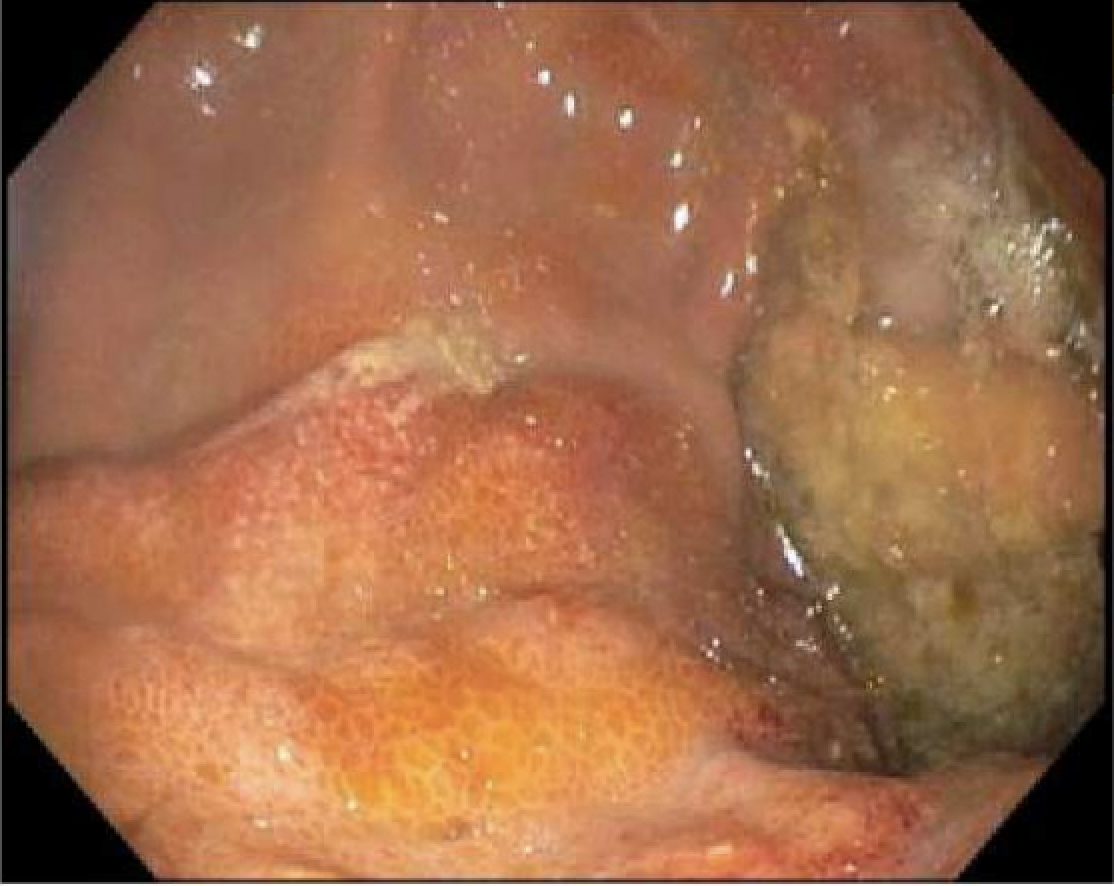
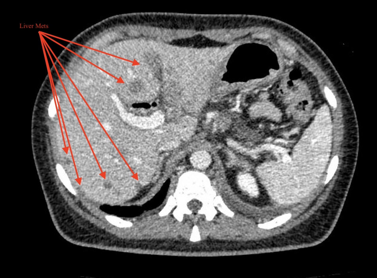
Methods: A 25-year-old female with no medical history presents for acutely worsening epigastric abdominal pain for 2 weeks. She had a recent admission to an outside hospital for similar symptoms and imaging demonstrating innumerable pulmonary nodules, numerous hypodense hepatic nodules, and multi-station lymphadenopathy, concerning for metastatic disease. Labs are notable for CA 125 elevated to 92 and CA 19-9 elevated to 1224. In addition, lipase was elevated, concerning for acute pancreatitis. Liver biopsy demonstrated poorly differentiated CK7 positive carcinoma consistent with suspected primaries as pancreatic, upper gastrointestinal (UGI), or lymphoma. EGD is notable for a nodular esophagus with random esophageal and gastric biopsies significant for poorly differentiated carcinoma favoring a UGI primary malignancy. Patient was started on FOLFOX prior to discharge to prevent further decompensation with an overall guarded prognosis due to tumor burden. Discussion: Linitis plastica has been historically difficult to diagnose as it typically infiltrates the submucosa and spares the mucosa. Thus, the typical superficial biopsies taken during EGD often produce inconclusive results (30-36%). High resolution CT with contrast is the gold standard for diagnosis; however, most cases are either discovered incidentally or at a late stage once extensive metastatic disease is present. In cases of extensive metastatic disease, palliative chemotherapy is favored to reduce overall malignant burden with total gastrectomy providing the only curative therapy. Surgical intervention increases the median survival from 3.6 months to 16.7 months. Total gastrectomy, however, is limited by several factors. Patients must have early stage, localized disease for total gastrectomy to be curative. Additionally, total gastrectomy is a major surgical intervention with a high risk of morbidity and mortality. Patients that survive the procedure are often plagued with chronic malnutrition. Overall, prognosis is guarded due to the advanced stage at which disease is typically diagnosed.
Figure: Poorly differentiated carcinoma
Figure: Numerous hypodense hepatic nodules
Disclosures: Parth Patel indicated no relevant financial relationships. Saatchi Kuwelker indicated no relevant financial relationships. Sherif Ibrahim indicated no relevant financial relationships. Hari Sayana: Boston Scientific – Consultant.
Parth Patel, MD, MBA1, Saatchi Kuwelker, MD1, Sherif Ibrahim, MD2, Hari Sayana, MD1. P4198 - Silent but Deadly: A Case of Linitus Plastica, ACG 2025 Annual Scientific Meeting Abstracts. Phoenix, AZ: American College of Gastroenterology.