Vijosh V. Kumar, MBBS, MD1, Sabu K. Gopi, MBBS, MD, DM1, Lijaya A, 2, Javed P, MBBS, MD, DM1, Kavitha Rangan, MBBS, MD, DM1, Vivek Kumar, MBBS, MD, DM1, Jaseen Ansari, MBBS, MD, DM1, Manas Jyoti Das, 2 1ASTER MIMS HOSPITAL KANNUR, Kannur, Kerala, India; 2NIT CALICUT, Kozhikode, Kerala, India Introduction: Helicobacter pylori (H. pylori) is well known for causing gastritis and gastric malignancy , necessitating accurate and timely diagnosis for effective management. While endoscopy allows direct visualization, advanced techniques like Narrow Band Imaging (NBI) with optical magnification enhance mucosal details associated with H. pylori infection. These subtle patterns, however, require expert interpretation, potentially leading to variability . This study aims to develop an artificial intelligence based system capable of accurately detecting and identifying H. pylori infection from endoscopic images obtained through Narrow Band Imaging. Methods: This prospective study was conducted at Aster MIMS Kannur, India, over a one-year period from March 2023 to March 2024.Sixty patients undergoing upper gastrointestinal endoscopy were recruited for the study after meeting the inclusion. The study cohort comprised two equal groups: 30 patients diagnosed as H. pylori-negative and 30 patients diagnosed as H. pylori-positive based on Rapid Urease test .High-resolution 4K NBI images with optical magnification were acquired and transferred for feature extraction at NIT Calicut. Features were extracted using Gabor Bank 2 filters combined with pixel values, excluding black pixels, resulting in a large feature set. Eight AI models (ANN, CNN, SVM on Gabor 1, Random Forest on Gabor Bank 2) were developed and their validation accuracy was assessed. Results: This study was a prospective investigation conducted at Aster MIMS Kannur from March 2023 to March 2024, involving 60 patients (30 H. pylori-positive and 30 H. pylori-negative). High-resolution 4K NBI images with optical magnification were acquired and transferred for feature extraction at NIT Calicut. Features were extracted using Gabor Bank 2 filters combined with pixel values, excluding black pixels, resulting in a large feature set. Eight AI models (ANN, CNN, SVM on Gabor 1, Random Forest on Gabor Bank 2) were developed and their validation accuracy was assessed. Discussion: This study identifies the potential of AI, particularly CNN and Random Forest, for H. pylori diagnosis using magnified NBI. The superior performance of the Random Forest on NBI images with Gabor Bank 2 features emphasizes the importance of both imaging modality and feature extraction. The write up used help with Gemini AI tool.
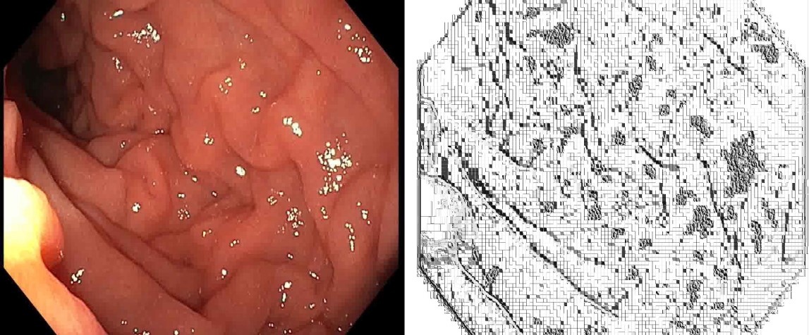
Figure: LBP extraction of an image
Disclosures: Vijosh V. Kumar indicated no relevant financial relationships. Sabu K. Gopi indicated no relevant financial relationships. Lijaya A indicated no relevant financial relationships. Javed P indicated no relevant financial relationships. Kavitha Rangan indicated no relevant financial relationships. Vivek Kumar indicated no relevant financial relationships. Jaseen Ansari indicated no relevant financial relationships. Manas Jyoti Das indicated no relevant financial relationships.
Vijosh V. Kumar, MBBS, MD1, Sabu K. Gopi, MBBS, MD, DM1, Lijaya A, 2, Javed P, MBBS, MD, DM1, Kavitha Rangan, MBBS, MD, DM1, Vivek Kumar, MBBS, MD, DM1, Jaseen Ansari, MBBS, MD, DM1, Manas Jyoti Das, 2. P4190 - Unlocking Microscopic Insights: AI-Driven Analysis of Magnified NBI for <i>H. pylori</i>, ACG 2025 Annual Scientific Meeting Abstracts. Phoenix, AZ: American College of Gastroenterology.

 photo")