University of Utah School of Medicine Salt Lake City, UT
Benjamin Gow-Lee, MD1, Jessica Schmitt, MD1, John Erikson Yap, MD, MBA, FACG2 1University of Utah School of Medicine, Salt Lake City, UT; 2University of Utah Health, Salt Lake City, UT Introduction: Adenomas are common precancerous lesions of the gastrointestinal tract, most frequently found in the colon. Small bowel adenomas are rare and typically associated with hereditary syndromes such as Familial Adenomatous Polyposis (FAP). This case presents a sporadic terminal ileal adenoma discovered incidentally in an immunocompromised patient without a known genetic predisposition.
Case Description/
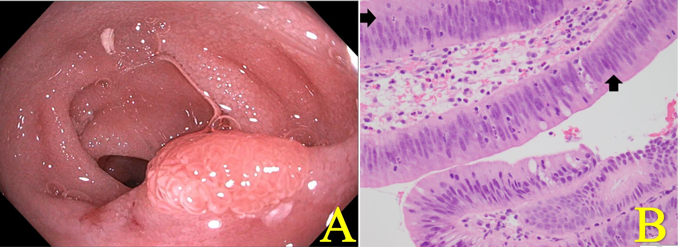
Methods: A 58-year-old man with relapsed multiple myeloma, recently treated with chimeric antigen receptor T-cell (CAR-T) therapy, presented with six weeks of intermittent, non-bloody watery diarrhea. Initial evaluation focused on infectious causes. A gastrointestinal PCR panel and Clostridioides difficile assay were unrevealing. Due to persistent symptoms, PET-CT imaging was performed and showed mild bowel wall thickening with increased radiotracer uptake from the rectum to the descending colon, raising concern for inflammation. Given the patient’s immunocompromised state and the possibility of CAR-T-related gastrointestinal toxicity, colonoscopy was pursued. The colon appeared endoscopically normal, but a small polyp was noted in the terminal ileum. Random colonic biopsies were histologically unremarkable. Histopathologic evaluation of the ileal lesion revealed a tubular adenoma (Images A & B), with a marked paucity of plasma cells and lymphocytes in the surrounding mucosa. The patient’s diarrhea resolved spontaneously in the days following the procedure. A pending viral PCR from the initial stool evaluation later returned positive for norovirus. Discussion: Small bowel adenomas represent less than 2% of gastrointestinal neoplasms and are most often found in the duodenum. Terminal ileal adenomas are especially rare and typically linked to hereditary syndromes, though sporadic cases can occur. These lesions are usually asymptomatic and often incidental but can progress to adenocarcinoma through a distinct molecular pathway. In this case, the ileal adenoma was likely unrelated to the patient’s symptoms. The observed mucosal immune depletion may reflect CAR-T-associated immunosuppression, potentially impairing IgA-mediated defenses and predisposing to enteric infection. This case underscores the value of thorough mucosal evaluation in immunocompromised patients with diarrhea, even when findings appear incidental.
Figure: A. Endoscopic image of terminal ileum showing adenoma. B. Hematoxylin and Eosin (H&E) stain demonstrating a high-power view of the terminal ileum with an adenoma. The penicillate picket fence nuclei (arrows) are typical of adenomas throughout the GI tract. Image was captured with a 40x objective.
Disclosures: Benjamin Gow-Lee indicated no relevant financial relationships. Jessica Schmitt indicated no relevant financial relationships. John Erikson Yap: Phathom Pharmaceutical – Speakers Bureau. Steris – Consultant.
Benjamin Gow-Lee, MD1, Jessica Schmitt, MD1, John Erikson Yap, MD, MBA, FACG2. P4141 - A Curious Coincidence: Sporadic Ileal Adenoma in an Immunocompromised Patient, ACG 2025 Annual Scientific Meeting Abstracts. Phoenix, AZ: American College of Gastroenterology.

.jpg "Benjamin Gow-Lee, MD (he/him/his) photo")