Julio C. Valencia-Manrique, MD1, Oyedotun Babajide, MD1, Gulce Karaca, MD2, Aria Khan, MD3, Ana Maritza Marulanda Prado, MD2, Dalia Mahmoud Hassan Elamin, MD2 1NYC Health + Hospitals/Metropolitan, New York, NY; 2NYC Health + Hospitals/Woodhull, Brooklyn, NY; 3NYC Health + Hospitals/Woodhull, Jamaica, NY Introduction: Pyogenic granuloma is a rare finding in the gastrointestinal tract, especially in the small bowel. It is a benign vascular lesion that can present with iron deficiency anemia or obscure gastrointestinal bleeding (GIB). The appearance may raise concern for malignancy, often prompting further workup with capsule endoscopy, deep enteroscopy, and imaging. Due to its rarity, it may not be immediately considered in the differential diagnosis. We present a case of jejunal pyogenic granuloma diagnosed during evaluation of unexplained iron deficiency anemia in a patient without overt GIB.
Case Description/
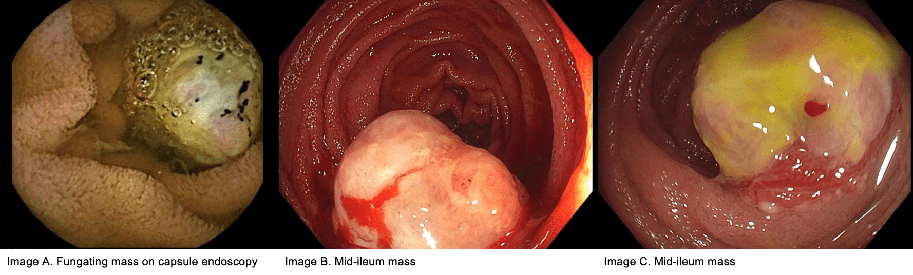
Methods: A 59-year-old female presented with fatigue and dizziness. Initial labs revealed hemoglobin of 7.0 g/dL. Iron studies confirmed iron deficiency anemia (IDA) without overt GIB. To evaluate for occult blood loss, EGD revealed chronic gastritis with intestinal metaplasia, without atrophic changes or stigmata of active bleeding. Colonoscopy showed a diminutive hyperplastic rectal polyp; the remainder of the exam was unremarkable. No clear bleeding source was found. Given the ongoing IDA and negative bidirectional endoscopy, capsule endoscopy was performed, revealing a medium-sized fungating lesion in the small bowel at 1 hour and 3 minutes. Push enteroscopy confirmed a 3 cm polypoid, fungating mass in the proximal jejunum, concerning for malignancy, without active bleeding. Biopsies were obtained, and the area around the lesion was tattooed. Cross-sectional imaging showed no local spread. Pathology demonstrated polypoid granulation tissue with superficial ulceration and was immunohistochemically positive for CD31, CD34, SMA, and Factor VIII consistent with a pyogenic granuloma. Discussion: Pyogenic granuloma (PG), or lobular capillary hemangioma, is a benign vascular lesion typically found on the skin or oral mucosa. In the GI tract, it is rare and may present as obscure GIB or iron deficiency anemia. Small bowel tumors account for < 5% of all GIB, making PG an uncommon but essential consideration. Fewer than 50 GI cases have been reported, with under 35 in the small intestine. In postmenopausal women with unexplained IDA and negative bidirectional endoscopy, capsule endoscopy is essential for small bowel evaluation. PGs may appear as polypoid or fungating masses, mimicking malignancy. Histology shows lobular capillary proliferation with granulation tissue. Immunostaining is typically positive for CD31 and CD34. Resection is recommended to confirm diagnosis and relieve symptoms.
Figure: Capsule endoscopy and push enteroscopy images
Disclosures: Julio Valencia-Manrique indicated no relevant financial relationships. Oyedotun Babajide indicated no relevant financial relationships. Gulce Karaca indicated no relevant financial relationships. Aria Khan indicated no relevant financial relationships. Ana Maritza Marulanda Prado indicated no relevant financial relationships. Dalia Mahmoud Hassan Elamin indicated no relevant financial relationships.
Julio C. Valencia-Manrique, MD1, Oyedotun Babajide, MD1, Gulce Karaca, MD2, Aria Khan, MD3, Ana Maritza Marulanda Prado, MD2, Dalia Mahmoud Hassan Elamin, MD2. P4138 - The Hidden Culprit: Occult Bleeding From a Small Bowel Pyogenic Granuloma, ACG 2025 Annual Scientific Meeting Abstracts. Phoenix, AZ: American College of Gastroenterology.