Wayne State University School of Medicine /John D. Dingell VA Medical Center Southfield, MI
Maria Fatima, MD1, Muni Hassan, BSc2, Fowzia Hassan, BSc2, Fadumo Mohammed, BSc2, Hameed Shanawaz, MD3, Muhammad Hashmi, MD4, Frederick Tiesenga, MD5 1Wayne State University School of Medicine /John D. Dingell VA Medical Center, Southfield, MI; 2Windsor University School of Medicine, Edmonton, AB, Canada; 3Detroit Medical Center/Wayne State University, Windsor, ON, Canada; 4Letterkenny University Hospital, Letterkenny, Donegal, Ireland; 5Suburban Surgery Center, Downers Grove, IL Introduction: Appendiceal mucinous neoplasms (AMNs) are rare epithelial tumors, accounting for less than 1% of appendectomy specimens. Their clinical presentation is often vague or incidental, but they carry a risk of serious complications, including pseudomyxoma peritonei. Low-grade appendiceal mucinous neoplasms (LAMNs), in particular, exhibit mucinous epithelial proliferation with low-grade cytologic atypia and potential for peritoneal spread. This case highlights the importance of early detection, surgical management, and surveillance of LAMNs due to their complex clinical course.
Case Description/
Methods: A 79-year-old male with a history of prostate cancer, hypertension, and arthritis presented with a year-long history of dull right lower quadrant abdominal pain and microscopic hematuria. Contrast-enhanced CT imaging revealed left nephrolithiasis and an incidental cystic lesion in the right lower quadrant, as well as two hepatic lesions. After urological evaluation cleared the renal concerns, the patient was referred to general surgery. He reported chronic constipation in addition to his abdominal discomfort.
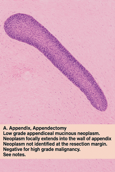
A laparoscopic appendectomy was performed, and histopathology revealed a distended appendix with a low-grade appendiceal mucinous neoplasm (LAMN) showing mural extension without high-grade features. Margins were negative for neoplastic involvement. Given the concern for peritoneal dissemination and indeterminate liver lesions, a right hemicolectomy and tru-cut liver biopsy were completed. Intraoperatively, no peritoneal spread was noted. Pathology showed a benign tubular adenoma in the colon and simple hepatic cysts. Postoperatively, the patient reported mild weight loss but was otherwise asymptomatic. He was advised to undergo regular colonoscopic surveillance. Discussion: LAMNs are often incidentally discovered but can have significant clinical implications if not fully resected. While appendectomy may be sufficient in confined cases, our decision to proceed with hemicolectomy was influenced by concern for potential spread and radiologic uncertainty. Although recent studies suggest hemicolectomy does not improve outcomes unless lymph nodes are involved, it remains common in select cases. Meticulous surgical technique is crucial to avoid rupture and pseudomyxoma peritonei. This case emphasizes the need for multidisciplinary evaluation and long-term surveillance, especially due to the potential for synchronous or metachronous neoplasia.
Figure: Figure 1. H&E-stained micrograph of the appendectomy specimen showing mucinous epithelium and mural extension consistent with low-grade appendiceal mucinous neoplasm (LAMN).
Disclosures: Maria Fatima indicated no relevant financial relationships. Muni Hassan indicated no relevant financial relationships. Fowzia Hassan indicated no relevant financial relationships. Fadumo Mohammed indicated no relevant financial relationships. Hameed Shanawaz indicated no relevant financial relationships. Muhammad Hashmi indicated no relevant financial relationships. Frederick Tiesenga indicated no relevant financial relationships.
Maria Fatima, MD1, Muni Hassan, BSc2, Fowzia Hassan, BSc2, Fadumo Mohammed, BSc2, Hameed Shanawaz, MD3, Muhammad Hashmi, MD4, Frederick Tiesenga, MD5. P4082 - Case of Massive Rare Mucinous Appendiceal Neoplasm With Peritoneal Dissemination, ACG 2025 Annual Scientific Meeting Abstracts. Phoenix, AZ: American College of Gastroenterology.

 photo")