Akhil Adla, DO1, Samuel Trenner, 2, Samantha Whitwell, MD2, Claudio Tombazzi, MD3 1University of Tennessee Health Science Center, Memphis, TN; 2UTHSC, Memphis, TN; 3VA Memphis Health Care, Memphis, TN Introduction: Extramedullary hematopoiesis (EMH) is a rarity in adults and occurs secondary to an underlying condition that causes chronic anemia. EMH typically occurs in the liver, spleen or lymph nodes and can present with non-specific symptoms depending on the site of EMH [3][5][6]. The non-specific nature of presentation as well as asymptomatic patients make diagnosis and investigation of EMH challenging. We present an unusual case of an asymptomatic 73-year-old man with a liver lesion that was found to be a site of extramedullary hematopoiesis which was initially concerning for malignancy.
Case Description/
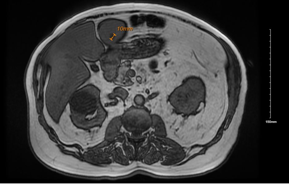
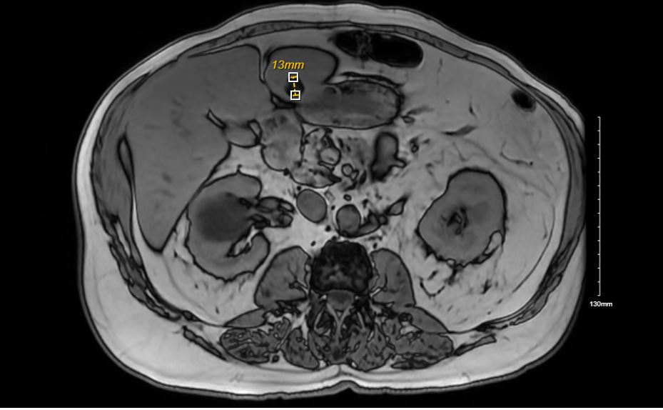
Methods: We present a 73-year-old man with past medical history of alcohol abuse and chronic back pain who presented with no symptoms but had an incidental finding of a 10mm hepatic nodule on MRI, initially concerning for focal area of fat versus malignancy. Physical exam was unremarkable without hepatomegaly. Patient’s labs were all within normal ranges with initial Alpha-fetoprotein (AFP) of 1.9. An MRI three months later showed a slight increase in the size and lesion was deemed to be LR3, demonstrating intermediate risk of hepatocellular carcinoma (HCC). Repeat imaging at 6 and 9 months from initial findings showed no significant changes. At the 15-month interval, there was concern for small cholangiocarcinoma due to increased size of the nodule to 13mm. Nuclear medicine tagged RBC study ruled out hemangioma. Biopsy of the nodule was performed which was negative for malignant cells but exhibited evidence for extramedullary hematopoiesis with trilineage hematologic cells, lymphocytes, adipocytes and benign hepatocytes. Imaging review showed a mildly decreased marrow signal observed in spine and pelvis, potentially representing hyperplasia or myeloproliferative disorder. Discussion: The report highlights the need for routine and close surveillance of liver lesions. The differential for liver lesions is large, and most of these conditions are treatable but early detection is essential for improved outcomes. In case of diagnostic concerns, tissue sampling is prudent choice because of the low levels of mortality and morbidity associated with liver biopsies. A systematic approach should be utilized to rule out life threatening conditions and enable patient to get treatment in a timely manner.
Figure: Figure A: Liver lesion initially measuring 10mm in 2022
Figure: Figure A: Liver lesion measuring 13mm in 2023 prompting a biopsy.
Disclosures: Akhil Adla indicated no relevant financial relationships. Samuel Trenner indicated no relevant financial relationships. Samantha Whitwell indicated no relevant financial relationships. Claudio Tombazzi indicated no relevant financial relationships.
Akhil Adla, DO1, Samuel Trenner, 2, Samantha Whitwell, MD2, Claudio Tombazzi, MD3. P3908 - Extramedullary Hematopoiesis in an Adult Presenting as an Asymptomatic Liver Lesion, ACG 2025 Annual Scientific Meeting Abstracts. Phoenix, AZ: American College of Gastroenterology.

 photo")