William Breaux, MD1, Lovekirat Dhaliwal, MD2, Saurabh Chawla, MD, FACG1 1Emory University School of Medicine, Atlanta, GA; 2Emory University Hospital, Atlanta, GA Introduction: Rectal cancer is among the most commonly diagnosed malignancies in the United States and typically presents with symptoms of rectal bleeding and altered bowel habits with presence of an intraluminal mass. The majority of colorectal adenocarcinomas arise from the mucosal lining of the bowel wall and are identified with direct visualization during colonoscopy. This case describes a rare presentation of colorectal adenocarcinoma manifesting as an extraluminal perirectal mass with no detectable intraluminal tumor seen on endoscopic imaging.
Case Description/
Methods: A 64-year-old male with a history of hypertension, hyperlipidemia, and peripheral artery disease initially presented with a 2-week history of buttock and back pain after a mechanical fall. Computed tomography (CT) imaging revealed an indeterminate 4.0 x 3.0 cm presacral soft tissue mass and lytic lesions within the left ilium concerning for metastatic disease. Further positive emission tomography (PET) CT showed a perirectal mass, left sacral lytic lesion, pulmonary nodules, and thoracic lymphadenopathy. Patient underwent colonoscopy showing multiple sessile polyps and extrinsic rectal compression without presence of an intraluminal mass in the rectum itself, which was biopsied and tattooed. Further imaging with endoscopic ultrasound showed a rectal wall mass arising from the muscularis propria, extending through the serosa. Fine needle biopsy showed poorly differentiated adenocarcinoma. The patient was started on FOLFOX with bevacizumab and continues to follow with oncology for treatment. Discussion: This case highlights a rare presentation of stage four rectal adenocarcinoma, in which the tumor was found extraluminally as a perirectal mass that was not visualized on intraluminal inspection. There is limited data describing rectal adenocarcinoma that originate outside the lumen, given that this histologic subtype typically arises from the mucosal lining. A prior case report noted presence of presacral adenocarcinoma, however the origin could not be determined due to tissue replacement given advanced disease. Extraluminal local recurrence of rectal cancer has been described in literature, but our patient had no prior history of colorectal cancer or report of prior polyp resection with subsequent findings of high-grade dysplasia of intramucosal cancer. This finding prompts further discussion regarding potential for atypical locations of rectal adenocarcinoma.
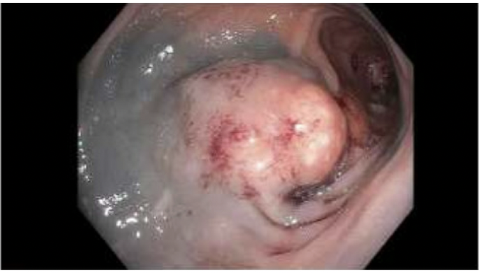
Figure: Image 1. Extrinsic compression 10cm from the anus on colonoscopy evaluation.
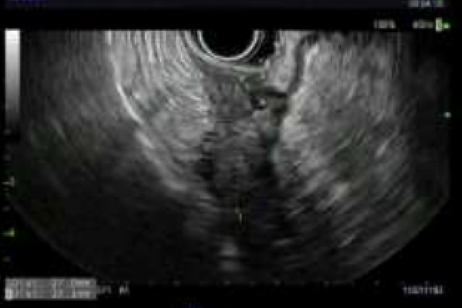
Figure: Image 2. Irregular hypoechoic mass arising from the muscularis propria and extending through the serosal layer at the mid rectum on endoscopic ultrasound evaluation.
Disclosures: William Breaux indicated no relevant financial relationships. Lovekirat Dhaliwal indicated no relevant financial relationships. Saurabh Chawla indicated no relevant financial relationships.
William Breaux, MD1, Lovekirat Dhaliwal, MD2, Saurabh Chawla, MD, FACG1. P4663 - Hidden in Plain Site: An Atypical Case of Extraluminal Rectal Adenocarcinoma, ACG 2025 Annual Scientific Meeting Abstracts. Phoenix, AZ: American College of Gastroenterology.