Abdelrahman Abdalla, 1, SriHari Mahadev, MD2, David Wan, MD3, Lillian F. Zhang, MD, PhD4, Adeyinka Charles. Adejumo, MD, MS, MSc5 1Weill Cornell Medicine, Doha, Ad Dawhah, Qatar; 2Weill Cornell Medicine, New York, NY; 3NewYork-Presbyterian Hospital/Weill Cornell Medical Center, New York, NY; 4NewYork-Presbyterian / Weill Cornell Medical Center, New York, NY; 5NewYork-Presbyterian Hospital/Weill Cornell Medical Center, Macon, GA Introduction: Esophageal injury due to spinal hardware is a rare but serious cause of dysphagia, which should be considered in patients with prior cervical spine surgery and radiation therapy. We describe a case of dysphagia due to cervical fusion hardware erosion into the esophageal lumen with ulcer formation in the context of prior Ewing’s sarcoma. The case illustrates how this complication may occur years and even decades after.
Case Description/
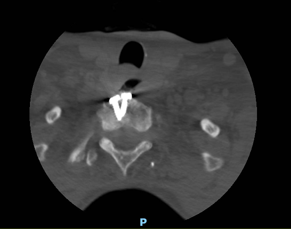
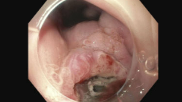
Methods: A 35-year-old man with a history of Ewing’s sarcoma treated at 8 years of age with C5–T2 corpectomy, anterior-posterior cervical fusion, and radiation therapy, presented with several weeks of progressive dysphagia. He described food impaction sensations, intermittent severe pain during meals, and a hoarse, deeper voice. Imaging demonstrated a prevertebral soft tissue mass spanning C5–T2, inseparable from the esophagus, concerning for residual or recurrent tumor. There were findings concerning for hardware erosion into the posterior esophageal wall, the cervical plate appeared to be partially exposed within the esophageal lumen. Esophagogastroduodenoscopy with endoscopic ultrasound revealed a transmural ulcer directly communicating with spinal fusion hardware. Biopsies of the ulcer showed active esophagitis with no evidence of malignancy. Biopsy of the surrounding tissue showed signs of chronic inflammation consistent with post-radiation fibrosis. Discussion: Plate erosion through the posterior esophageal wall is a late complication of cervical spine surgery that may present years or even decades after cervical fusion surgery. This complication has been documented in case series, often presenting with dysphagia, hoarseness, and perforation. What makes our case unique is the fact that this complication manifested for the first time 27 years after surgery, making this the longest reported interval between the surgery and esophageal erosion. Previously, the longest documented interval between anterior cervical spine surgery and the first manifestation of esophageal erosion by spinal hardware was 22 years. Literature suggests that chronic mechanical stress from cervical hardware as well as local tissue ischemia and fibrosis from radiation therapy may weaken tissue integrity and contribute to gradual erosion of the esophageal wall. Despite these associations, the inciting event leading to such a late-onset complication is not clearly identifiable, highlighting the unpredictable nature of this rare but serious outcome.
Figure: CT of the Cervical Spine
Figure: EGD Showing Ulceration in the Upper Third of the Esophagus
Disclosures: Abdelrahman Abdalla indicated no relevant financial relationships. SriHari Mahadev: Boston scientific – Consultant. Conmed – Consultant. David Wan: Medtronic – Data Monitoring Committee. Lillian Zhang indicated no relevant financial relationships. Adeyinka Adejumo indicated no relevant financial relationships.
Abdelrahman Abdalla, 1, SriHari Mahadev, MD2, David Wan, MD3, Lillian F. Zhang, MD, PhD4, Adeyinka Charles. Adejumo, MD, MS, MSc5. P5043 - Delayed Erosion of an Anterior Cervical Spine Plate Into the Esophagus 27 Years After Surgery and Radiotherapy, ACG 2025 Annual Scientific Meeting Abstracts. Phoenix, AZ: American College of Gastroenterology.