Andrew Guido, MMS1, Zubair Khan, MD2, Edwardo Verzola, MD2, Saad Zinad, MD, MPH2 1Mercy Hospital Jefferson, Baldwinsville, NY; 2Mercy Hospital Jefferson, Festus, MO Introduction: Gastrointestinal stromal tumors (GISTs) are rare mesenchymal neoplasms originating from the interstitial cells of Cajal, accounting for 1–2% of all primary gastrointestinal (GI) tumors. They occur most commonly in the stomach, followed by the small bowel. Of all small bowel GISTS, ~ 10% arise in the jejunum, and ~ 25% present with GI bleeding; sporadic multifocal cases of GISTs are rare. Diagnosis is based on imaging, histopathology, and immunohistochemical (IHC) staining, notably for CD117. We present a case of bleeding sporadic multifocal jejunal GIST to highlight diagnostic challenges and discuss management in the context of a literature review.
Case Description/
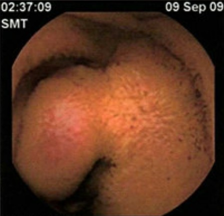
Methods: A 56-year-old female presented for two months of abdominal pain, melena, maroon-colored stool, nausea, and fatigue. Relevant medical history included two remote episodes of GI bleeding with normal esophagogastroduodenoscopies (EGDs) and colonoscopies. Capsule endoscopy at that time revealed a jejunal nonbleeding lesion (Fig. 1). Her history included a Cesarean section and smoking without liver disease, NSAID, alcohol, or illicit drug use.
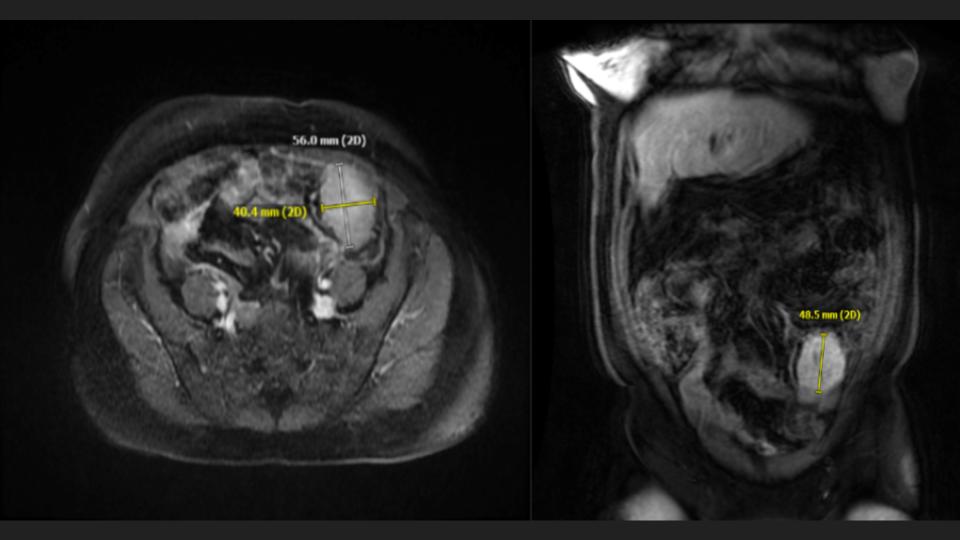
On admission, she was afebrile with a blood pressure of 100/60, heart rate of 102, and respiratory rate of 18. Physical exam revealed periumbilical tenderness, hyperactive bowel sounds, external hemorrhoids, and black tarry stool. Bloodwork showed severe anemia that improved with fluid resuscitation and blood transfusion. Computed tomography revealed a potential small bowel mass, confirmed by magnetic resonance enterography (Fig. 2). Repeat EGD and colonoscopy were negative for a mass or bleeding source.
Ongoing hemoglobin drops and symptoms led to an exploratory laparotomy, resulting in resection of a multifocal mid-to-distal jejunal mass. IHC stains were positive for CD117 and other GIST-associated markers, diagnosing multifocal GIST. She recovered well and was discharged on imatinib therapy. At one-year follow-up, she remained symptom-free and in good health. Discussion: A literature review identified two reports of Neurofibromatosis type 1 (NF1)–associated bleeding multifocal jejunal GISTs. To our knowledge, this is the first reported case of a sporadic bleeding multifocal jejunal GIST. The importance of GIST inclusion in the differential diagnosis of obscure GI bleeding, particularly when endoscopic evaluation is negative, is crucial. Early recognition and intervention are critical; delayed diagnosis may lead to increased morbidity and mortality—especially in resource-limited settings.
Figure: Fig. 1. Capsule endoscopy showing a nonbleeding potential nonbleeding polyp versus ulcer in the jejunum.
Figure: Fig. 2. Magnetic resonance enterography showing a 5.6 cm small bowel mass
Disclosures: Andrew Guido indicated no relevant financial relationships. Zubair Khan indicated no relevant financial relationships. Edwardo Verzola indicated no relevant financial relationships. Saad Zinad indicated no relevant financial relationships.
Andrew Guido, MMS1, Zubair Khan, MD2, Edwardo Verzola, MD2, Saad Zinad, MD, MPH2. P5269 - Multifocal Jejunal GIST Presenting With Gastrointestinal Bleeding: A Case Report and Literature Review, ACG 2025 Annual Scientific Meeting Abstracts. Phoenix, AZ: American College of Gastroenterology.