Rao Afzal, MD, Hafsa Khan, DO, Mohammed Y. Youssef, MD, Jaison John, MD Hunt Regional Medical Center, Greenville, TX Introduction: Upper gastrointestinal bleeding (UGIB) is a life-threatening medical emergency with mortality rates of 5%-10%. It occurs most commonly due to esophageal varices or peptic ulcer disease. Cystogastrostomy stents have been increasingly utilized to manage pancreatic pseudocyst drainage. This case highlights a massive UGIB secondary to cystogastrostomy stent mucosal erosion.
Case Description/
Methods: A 64-year-old male with past medical history of a prior pancreatic pseudocyst with cystogastrostomy stent, presented to the ED 3 months later with hematemesis, epigastric pain, and a clinical picture consistent with shock. CT scan of the abdomen showed signs of mucosal erosion in the gastric wall. An emergent upper endoscopy was performed revealing large amounts of blood clots obscuring the mucosal visibility. The patient’s cystogastrostomy stent was seen covered with large amounts of blood, and appeared to be dislodged. He was transferred to a tertiary hospital and underwent IR embolization, and removal of the stent.
Discussion: In conclusion, this case highlights a rare but life-threatening complication of LAMS placement—rupture of a gastric vessel due to stent erosion. Gastrointestinal bleeding from such vascular injury can be difficult to diagnose, especially when initial endoscopic evaluations are inconclusive. A high index of suspicion is essential once common sources of bleeding are ruled out. Early angiographic evaluation and embolization are critical for effective management and can significantly improve outcomes. Based on current literature and our experience, it is advisable to perform follow-up cross-sectional imaging within 3 to 4 weeks post-LAMS placement and to consider early removal of the stent once cyst resolution is achieved, to minimize the risk of vascular erosion and hemorrhage.
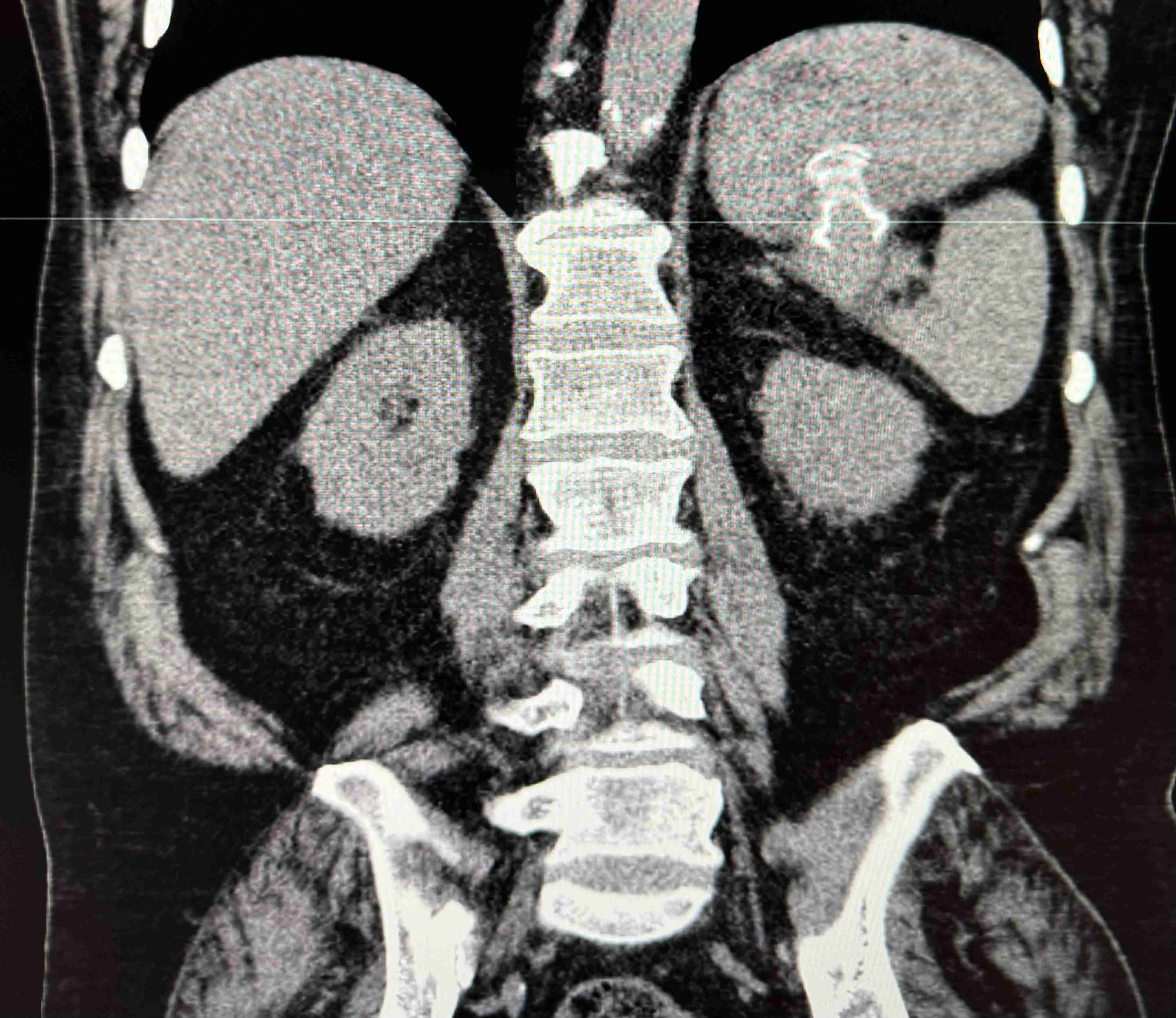
Figure: Non-contrast CT of the abdomen demonstrating focal gastric wall thickening surrounding the lumen-apposing metal stent (LAMS), representing gastric wall edema and hemorrhage. Presence of high-attenuation fluid within the gastric lumen is consistent with intraluminal hemorrhage. Additional focus of gas interposed between the stomach and spleen consistent with focal perforation of the gastric wall.
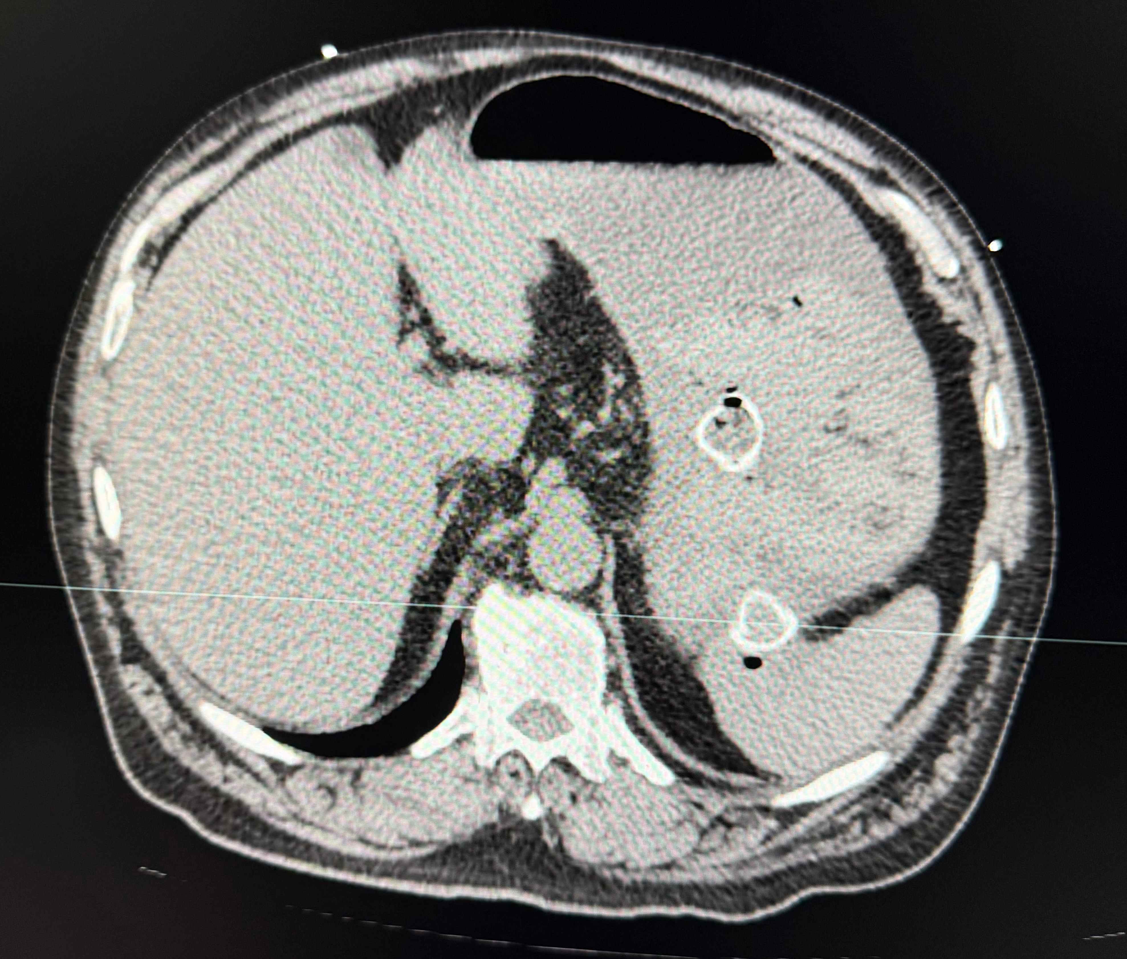
Figure: Focus of gas interposed between the stomach and spleen with adjacent fluid consistent with focal gastric wall erosion and localized hemorrhage.
Disclosures: Rao Afzal indicated no relevant financial relationships. Hafsa Khan indicated no relevant financial relationships. Mohammed Y. Youssef indicated no relevant financial relationships. Jaison John indicated no relevant financial relationships.
Rao Afzal, MD, Hafsa Khan, DO, Mohammed Y. Youssef, MD, Jaison John, MD. P5265 - From Cure to Crises: A Rare Case of Massive Upper GI Bleed Caused by Lumen-Apposing Metal Stent, ACG 2025 Annual Scientific Meeting Abstracts. Phoenix, AZ: American College of Gastroenterology.