University of Florida College of Medicine Gainesville, FL
Abdillahi Ahmed, MD1, Lindsey A. Creech, DO, MBA, MPH2, Benjamin Kaminski, MD1, Aleksey Novikov, MD1 1University of Florida College of Medicine, Gainesville, FL; 2University of Florida, Gainesville, FL Introduction: Mesothelioma is a rare cancer that typically affects the pleural mesothelium; however, pericardial involvement is exceptionally rare. We report a case of primary pericardial epithelioid mesothelioma in an adult where, despite initial consideration of surgical sampling, a diagnosis was achieved via endoscopic ultrasound (EUS)-guided biopsy. This case highlights the potential of EUS-guided sampling in the thorax.
Case Description/
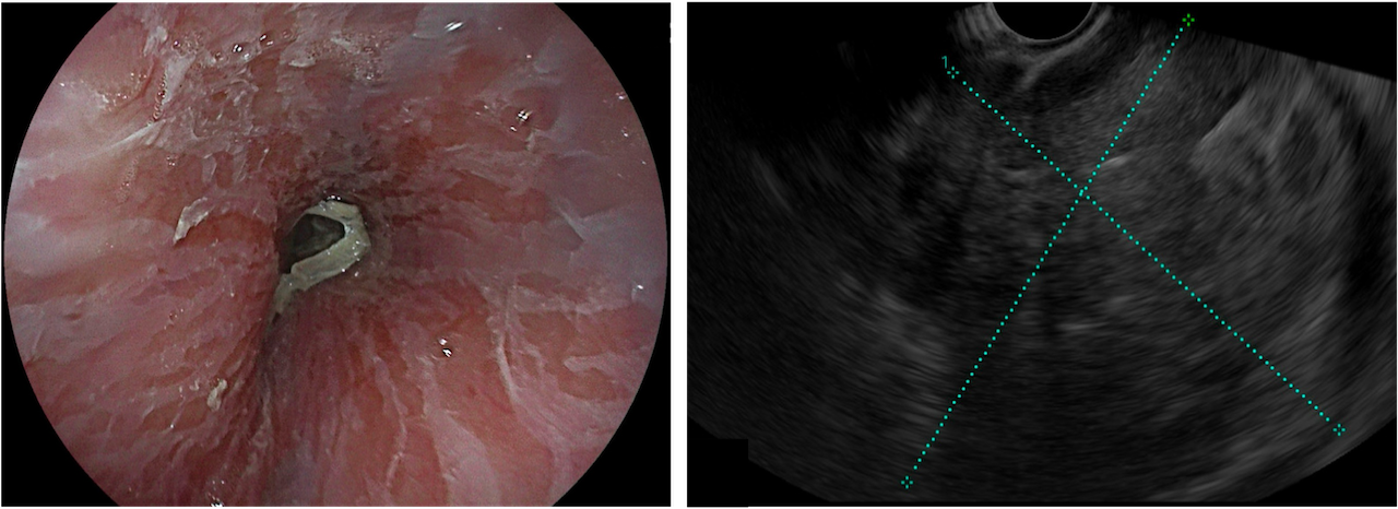
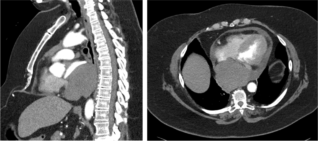
Methods: A 71-year-old female with atrial fibrillation and gastroesophageal reflux disease presented to the emergency department with several months of productive cough and acutely progressive shortness of breath. A computed tomography (CT) pulmonary angiogram revealed a 7.8 × 8.3 × 8.4 cm posterior mediastinal mass indistinguishable from the anterior esophagus, which effaced and compressed the left atrium, along with a moderate pericardial effusion measuring 14.7 mm in maximal thickness. Cardiac magnetic resonance imaging depicted similar mass characteristics with high vascularity and late gadolinium enhancement in the pericardium, suggesting infiltration. An upper endoscopy revealed diffuse mucosal sloughing and external compression of the middle esophagus. A fine needle biopsy was performed at the location of a hypoechoic 67 × 63 mm lesion visualized with endoscopic ultrasound (EUS) at 30 cm from the incisors. Tumor pathology showed discohesive epithelioid cells with eosinophilic cytoplasm infiltrating the underlying stroma. These cells were immunoreactive for calretinin, pancytokeratin, and cytokeratin 5/6, but negative for TTF-1. The patient was diagnosed with stage cT4N0M0 (IIIB) primary pericardial epithelioid mesothelioma. Notably, her social history was negative for tobacco use and overt asbestos exposure. She was not a candidate for surgical resection or radiotherapy due to pericardial tumor invasion. After six cycles of carboplatin and pemetrexed, five of which included bevacizumab, a CT chest showed the tumor size decreased to 5.9 × 5.9 × 5.4 cm and the pericardial effusion resolved. Discussion: This case demonstrates that EUS-FNB can be used as a minimally invasive method for sampling cardiac lesions in patients with thoracic malignancies. To our knowledge, EUS-FNB sampling of a cardiac lesion has only been reported once before.1 Therefore, this represents the second case described in the literature.
Haja A, Lakhtakia S, Sekaran A, et al. Cardiac sarcoma diagnosed with EUS-FNB. Endosc Int Open. 2021;9(2):E152-E153. doi:10.1055/a-1268-7675
Figure: Figure 1. Upper endoscopic image showing sloughing of the esophageal mucosa (left) and endoscopic ultrasound image depicting a 67 × 63 mm lesion located 30 cm from the incisors (right).
Figure: Figure 2. CT pulmonary angiogram in the late pulmonary arterial phase, shown in soft tissue window, demonstrates a 7.8 × 8.3 × 8.4 cm posterior mediastinal mass in the sagittal (left) and axial (right) planes.
Disclosures: Abdillahi Ahmed indicated no relevant financial relationships. Lindsey Creech indicated no relevant financial relationships. Benjamin Kaminski indicated no relevant financial relationships. Aleksey Novikov indicated no relevant financial relationships.
Abdillahi Ahmed, MD1, Lindsey A. Creech, DO, MBA, MPH2, Benjamin Kaminski, MD1, Aleksey Novikov, MD1. P5722 - Primary Pericardial Mesothelioma Diagnosed With Endoscopic Ultrasound Fine Needle Biopsy, ACG 2025 Annual Scientific Meeting Abstracts. Phoenix, AZ: American College of Gastroenterology.