Hasham Masood Qureshi, MD, Deepthi Devagudi, MD, Sana Khan, MD, Maheshwari N. Siddaraju, MD Adventist Health System, Hanford, CA Introduction: Coccidiomycosis is a dimorphic fungus endemic to Southwestern US and Northern Mexico which commonly causes a self-limiting lung disease. This report highlights an uncommon extra-pulmonary manifestation in peritoneum, with only 36 cases reported in literature.
Case Description/
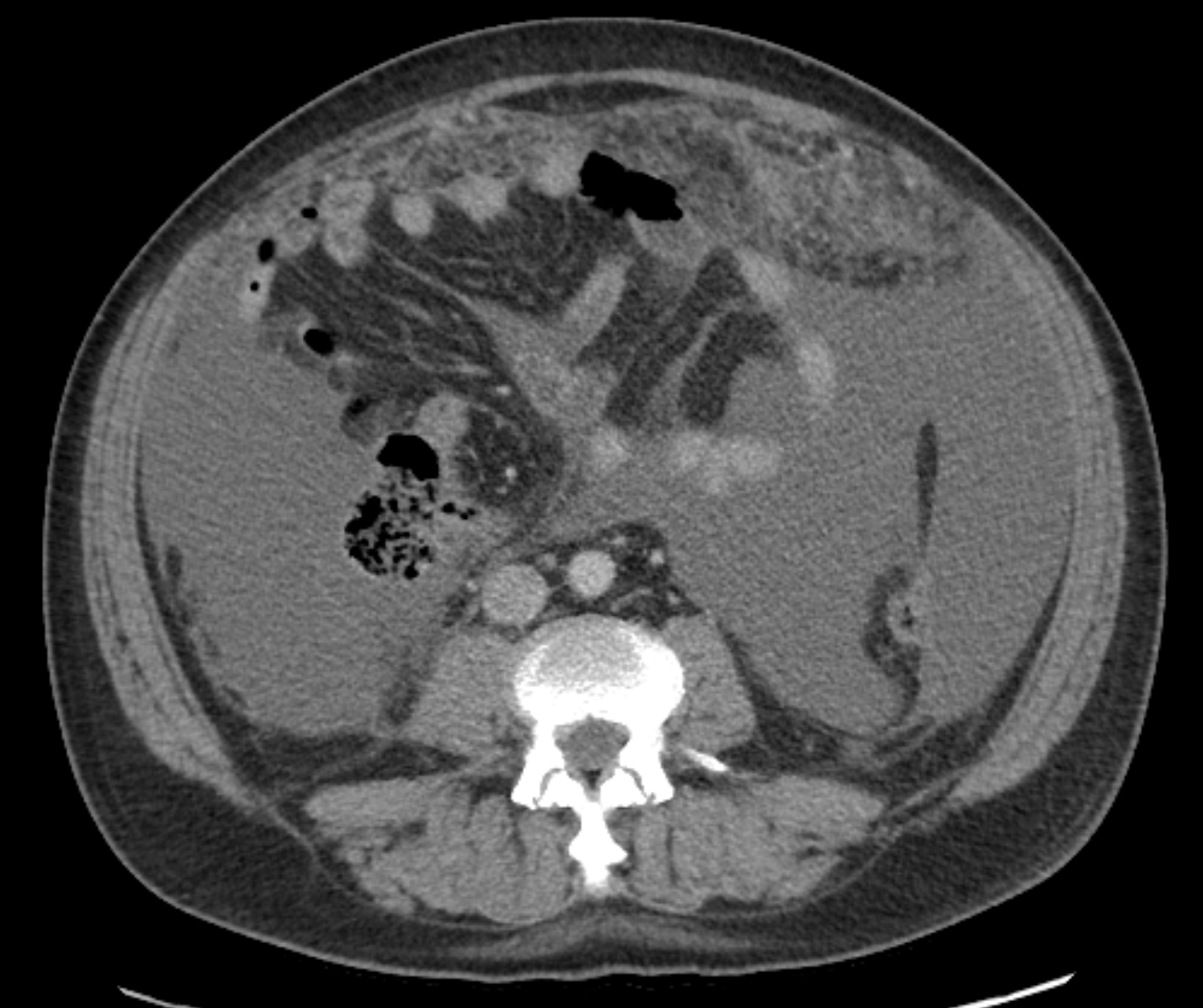
Methods: A 58 year old male from California presented to the hospital with progressive abdominal distention and nausea for 2 weeks. He also reported intermittent fever, night sweats and appetite changes for 3 months, along with two facial lesions that did not respond to trimethoprim-sulfamethoxazole 10-day course. CT imaging revealed extensive ascites and omental infiltration, while a chest CT showed mediastinal and hilar lymphadenopathy. Initially suspected to have an abdominal malignancy, an omental biopsy was ordered. Ascitic fluid analysis showed 2,100 WBCs, with 37% eosinophils and 51% lymphocytes. The Serum Ascites Albumin Gradient (SAAG) was < 1.1. Colonoscopy and esophagogastroduodenoscopy were unremarkable. Further testing, including alpha-fetoprotein, carbohydrate antigen 19-9, and carcinoembryonic antigen, were unremarkable, and the tuberculosis QuantiFERON was negative. The IgE level was 1,088 IU/mL. Given the symptoms of systemic fatigue, fever, cutaneous lesions, and now abdominal distension, coccidioidomycosis was considered. A repeat diagnostic paracentesis showed positive fungal block, abundant eosinophils presence, spherules and endospores consistent with coccidioides. The patient was started on fluconazole 800 mg daily and discharged to home with the plan to follow up with infectious disease specialist. Discussion: Coccidioides is a genus of dimorphic fungi that exist as mycelia or spherules. The infectious particles of Coccidioides are deposited in the lungs and transform into spherules, which may leave the lung to set up an extra-pulmonary infection in mostly immunocompromised patients. In patients with ascites due to disseminated cocci, the diagnosis can be based on standard peritoneal fluid analysis. Ascites in this context may cause significant morbidity if not diagnosed and treated promptly. This case highlights that disseminated coccidioidomycosis can occur even in immunocompetent individuals, although risk factors, such as ethnicity and geographic location, increase susceptibility. Rapid diagnosis via cytology can facilitate timely anti-fungal therapy. Recognition of systemic symptoms and atypical presentations in endemic regions is essential for effectively managing and treating this infection.
Figure: Omental thickening and ascites
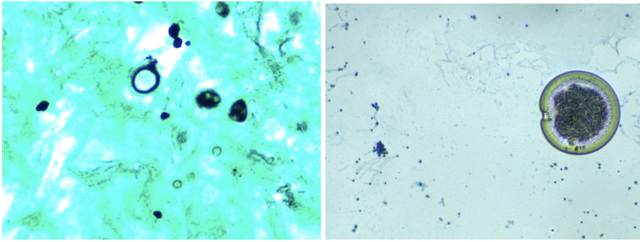
Figure: Coccidiomycosis endospore and GMS stain
Disclosures: Hasham Masood Qureshi indicated no relevant financial relationships. Deepthi Devagudi indicated no relevant financial relationships. Sana Khan indicated no relevant financial relationships. Maheshwari Siddaraju indicated no relevant financial relationships.
Hasham Masood Qureshi, MD, Deepthi Devagudi, MD, Sana Khan, MD, Maheshwari N. Siddaraju, MD. P5629 - Ascites-Suspected Carcinomatosis With a Surprising Finding: A Story of Disseminated Coccidioidomycosis, ACG 2025 Annual Scientific Meeting Abstracts. Phoenix, AZ: American College of Gastroenterology.

 photo")