Shamayel Safdar, MBBS, Erica Jensen, DO, Justin Bilello, MD, Rabbia Irfan, MBBS, Mohsin Chundrigar, MBBS, Seth Richter, MD, FACG, Stephen Hasak, MD, MPH Albany Medical Center, Albany, NY Introduction: Cytomegalovirus (CMV) is a dsDNA virus belonging to the Herpesviridae family. While CMV infection is typically asymptomatic in immunocompetent individuals, it can cause significant morbidity and mortality in immunocompromised individuals. We report a rare case of CMV jejunitis in a kidney transplant recipient presenting with chronic diarrhea and small bowel obstruction (SBO), mimicking Crohn’s Disease on imaging and endoscopy.
Case Description/
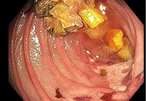
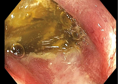
Methods: 46-year-old male with history of ESRD status post deceased donor kidney transplant on mycophenolate, belatacept, and tacrolimus presented with worsening abdominal pain and diarrhea for 6 months. Symptoms persisted after discontinuation of mycophenolate. Initial imaging and serum CMV PCR testing were unremarkable. Repeat computed tomography of abdomen and pelvis (CTAP) revealed distal SBO and mesenteric lymphadenopathy, normal esophagogastroduodenoscopy and colonoscopy with negative biopsies, and elevated fecal calprotectin 467 µg/g, concerning for inflammatory bowel disease (IBD). Magnetic resonance enterography revealed 8cm segmental distal ileitis. Crohn’s Disease was suspected. He was readmitted with worsening symptoms. CTAP demonstrated partial SBO with a transition point in the jejunum and small bowel Enteroscopy revealed severe ulceration and stenosis in the mid jejunum, associated with a large amount of food that could not pass. Biopsies were obtained. Crohn’s Disease remained the leading differential, and the patient was empirically started on prednisone while awaiting histology.Surprisingly, biopsy showed Active CMV with inclusion bodies appearing within infected cells. Prednisone was discontinued, intravenous ganciclovir was initiated for 10 days, followed by a prolonged oral course. The patient’s symptoms resolved and he was discharged. Discussion: CMV enteritis is exceedingly rare and presents a diagnostic challenge because the non-specific clinical presentation overlaps with a variety of conditions, including IBD, drug-induced enteritis, and bacterial or parasitic infections. Radiologic and endoscopic findings may mimic Crohn’s Disease, including mucosal ulceration, strictures, and mesenteric lymphadenopathy. Negative blood CMV PCR, as seen in this case, is common in isolated gastrointestinal involvement and should not exclude CMV from the differential. Misdiagnosis may result in inappropriate treatment, such as immunosuppressive corticosteroids, which can exacerbate viral infection and worsen outcomes.
Figure: Severe Ulceration and Stenosis in the mid jejunum
Figure: Large amount of food that could not pass secondary to stenosis
Disclosures: Shamayel Safdar indicated no relevant financial relationships. Erica Jensen indicated no relevant financial relationships. Justin Bilello indicated no relevant financial relationships. Rabbia Irfan indicated no relevant financial relationships. Mohsin Chundrigar indicated no relevant financial relationships. Seth Richter indicated no relevant financial relationships. Stephen Hasak: Castle Biosciences – Speakers Bureau. Conmed – Consultant.
Shamayel Safdar, MBBS, Erica Jensen, DO, Justin Bilello, MD, Rabbia Irfan, MBBS, Mohsin Chundrigar, MBBS, Seth Richter, MD, FACG, Stephen Hasak, MD, MPH. P5626 - Rare Presentation of CMV Jejunitis Masquerading as Crohn’s Disease: A Case Report, ACG 2025 Annual Scientific Meeting Abstracts. Phoenix, AZ: American College of Gastroenterology.


 photo")