P5518 - A Macroscopic Case of Microscopic Colitis? - A Case Demonstrating Endoscopic Findings in Microscopic Colitis and Subtypes to Consider Beyond Lymphocytic and Collagenous Variants
Tenzin Tseky, DO, Paige Robinson, DO, Brian Sowka, DO, Brendan Gregg, DO, Jose Russe-Russe, MD, Bruno Mazza, MD, Gary Valvano, DO Arnot Ogden Medical Center, Elmira, NY Introduction: Microscopic colitis (MC) is an entity of inflammatory bowel disease (IBD) associated with chronic watery diarrhea, normal colonoscopy, and traditionally classified into lymphocytic (LC) vs. collagenous (CC) subtypes. We present a case of patient with chronic diarrhea whose colonoscopy was notable for diffuse mild colonic mucosal changes with non-diagnostic histologic features on random biopsies suggestive of MC vs. traditional IBD in correct clinical setting per pathologist. This was treated as MC with budesonide with excellent response. This case highlights the importance of recognizing that MC can have macroscopic changes and that histologic diagnosis need not be limited to the two distinct subtypes.
Case Description/
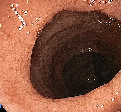
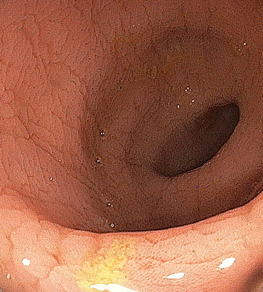
Methods: A 75-year-old female underwent colonoscopy for history of chronic loose stools with low fecal pancreatic elastase (otherwise unremarkable serological and stool studies). Colonoscopy was notable for diffuse mild colonic cobblestoning pattern and loss of vascularity. Random biopsies from right and left colon demonstrated non-specific histologic changes (features of erosion, mild acute cryptitis, mild crypt distortion, focal granulomatous changes) non-diagnostic for, and hence suggestive of either MC vs. traditional IBD in the correct clinical setting per pathologist report. As the patient lacked alarm features of traditional IBD, this was treated as MC and she was started on budesonide course with almost immediate response and successful taper. Discussion: MC has been characterized by the triad of chronic watery diarrhea (notable triggers include NSAIDs, SSRIs, tobacco, co-morbid Celiac Disease), normal colonoscopy, and classic histologic findings of CC vs LC. Initial treatment is aimed at removing identifiable triggers and/or budesonide course. Our case illustrates the importance of challenging these two classical notions regarding this disease entity: 1) MC can feature macroscopic changes. Particularly described in the literature are patterns associated with CC (pseudomembranes; mucosal vascular pattern alterations, nodularity, and defects). Our case noted endoscopic features of mild cobblestoning and loss of vascular pattern 2) There is an incomplete variant of MC by histology. Our case featured histologic changes not fitting into CC vs. LC 3) Despite these deviating from traditional notions of MC on both the macroscopic and microscopic levels, usual treatment protocol should be followed if clinical suspicion for MC is high, with expectations for excellent prognosis.
Figure: Diffuse Mild Cobblestoning and Loss of Vascularity noted on Colonoscopy
Figure: Diffuse Mild Cobblestoning and Loss of Vascularity noted on Colonoscopy
Disclosures: Tenzin Tseky indicated no relevant financial relationships. Paige Robinson indicated no relevant financial relationships. Brian Sowka indicated no relevant financial relationships. Brendan Gregg indicated no relevant financial relationships. Jose Russe-Russe indicated no relevant financial relationships. Bruno Mazza indicated no relevant financial relationships. Gary Valvano indicated no relevant financial relationships.
Tenzin Tseky, DO, Paige Robinson, DO, Brian Sowka, DO, Brendan Gregg, DO, Jose Russe-Russe, MD, Bruno Mazza, MD, Gary Valvano, DO. P5518 - A Macroscopic Case of Microscopic Colitis? - A Case Demonstrating Endoscopic Findings in Microscopic Colitis and Subtypes to Consider Beyond Lymphocytic and Collagenous Variants, ACG 2025 Annual Scientific Meeting Abstracts. Phoenix, AZ: American College of Gastroenterology.