Rhema Dadala, BS, Benjamin Richter, MD, Shivani Patel, MD, Raquel Olivo Salcedo, MD, Anmol Mittal, MD, Steven Krawitz, MD, Kaveh Hajifathalian, MD, Ahmed Al-Khazraji, MD Rutgers New Jersey Medical School, Newark, NJ Introduction: Heterotopic pancreatic tissue (HPT) is believed to form by pancreatic tissue from the dorsal and ventral buds adhering to the duodenum during development or from in situ differentiation of endodermal tissue. HPT is a rare finding, and it occurs most frequently in the gastric antrum (27%), duodenum (26%), and jejunum (20%). It is also found on 1 % of autopsies. HPT in the stomach is typically associated with large, life-threatening hemorrhage—distinct from the mild, indolent gastrointestinal bleeding more commonly seen with jejunal HPT. Here, we report a rare case of heterotopic pancreas in the jejunum (HPJ) causing massive GI bleeding.
Case Description/
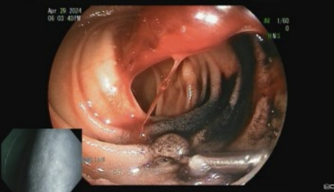
Methods: A 46 year old Male with a past medical history of HTN, HF-EF (30-35%), ESRD, DM, and anemia of chronic disease presented with weakness, shortness of breath, and melena with a hemoglobin of 3.2 g/dL. Patient underwent cross sectional imaging CT the abdomen and pelvis, EGD, and colonoscopy, none of which revealed a definitive source of bleeding. A capsule endoscopy identified bleeding in the jejunum but did not reveal the source. He remained in the hospital for ten days with ongoing large volume melana necessitating 16 units of pRBC over this period. Patient then underwent double balloon enteroscopy demonstrates evidence of small bowel bleeding with a medium sized blood clot in the proximal jejunum. This was tattooed and clipped for location. Tc-99m-labeled-RBC scan revealed focal abnormal radiotracer accumulation in the proximal small bowel. Patient underwent exploratory laparotomy and jejunal resection at HPJ site. Pathology confirmed HPT and congested blood vessels. Patient was discharged without further bleeding. Discussion: This is a rare case of HPT that presented with massive, small bowel bleeding. The proposed pathogenesis involves chronic inflammation within the ectopic pancreatic tissue, leading to the release of proteolytic enzymes such as trypsin and elastase. These enzymes can damage local blood vessels, resulting in hemorrhagic suffusion. This case highlights the importance of considering ectopic pancreatic tissue—alongside more common causes like arteriovenous malformations and ulcerations—as a rare but significant etiology of obscure small bowel bleeding.
Figure: Endoscopic imaging of HPT in proximal jejunum
Disclosures: Rhema Dadala indicated no relevant financial relationships. Benjamin Richter indicated no relevant financial relationships. Shivani Patel indicated no relevant financial relationships. Raquel Olivo Salcedo indicated no relevant financial relationships. Anmol Mittal indicated no relevant financial relationships. Steven Krawitz indicated no relevant financial relationships. Kaveh Hajifathalian indicated no relevant financial relationships. Ahmed Al-Khazraji indicated no relevant financial relationships.
Rhema Dadala, BS, Benjamin Richter, MD, Shivani Patel, MD, Raquel Olivo Salcedo, MD, Anmol Mittal, MD, Steven Krawitz, MD, Kaveh Hajifathalian, MD, Ahmed Al-Khazraji, MD. P5289 - A Rare Culprit for Small Bowel Hemorrhage: Ectopic Pancreatic Tissue in the Jejunum, ACG 2025 Annual Scientific Meeting Abstracts. Phoenix, AZ: American College of Gastroenterology.