NYC Health + Hospitals/South Brooklyn Health Brooklyn, NY
Sara Sadeghi, MD, Nicholas Bulba, BA, Akil Olliverrie, MD, Joshua Diaz, MD, Mohammad Chowdhury, MD, John Trillo, MD NYC Health + Hospitals/South Brooklyn Health, Brooklyn, NY Introduction: Gastrointestinal lipomas are very rare, only occurring in less than 1% of all gastrointestinal tumors and can range from asymptomatic to causing abdominal pain, dyspepsia, gastric outlet obstruction, and hemorrhage when over 2 cm. We present a case of an ulcerating gastric lipoma measuring 6 cm leading to UGIB. This case also highlights a successful treatment plan using endoscopic removal, bypassing the need for intensive gastric surgery.
Case Description/
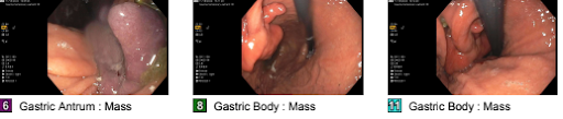
Methods: This is a case of a 72-year-old Russian female patient who presented to the hospital with exertional chest pain and was admitted to CCU for NSTEMI which later converted to STEMI. The post-cardiac catheterization course was complicated by acute UGIB. GI was consulted and the patient underwent endoscopy (EGD) which showed no active bleeding and a large (6 cm), pedunculated polypoid mass with ulceration on the posterior wall of the stomach. Biopsies were taken with cold forceps for histology to rule out malignancy; however, samples were insufficient. After 5 weeks, EGD was repeated and the mass was removed endoscopically. The pathology result was conclusive for well-circumscribed mature adipose tissue in the submucosa, compatible with lipoma and negative for dysplasia or carcinoma. Discussion: Lipomas consist of mature adipose cells surrounded by a thin fibrous capsule that usually occur superficially in the subcutaneous tissue and rarely in visceral organs. Gastric lipomas have no malignant potential and tend to arise in the antrum of the stomach. Asymptomatic gastric lipomas tend to be diagnosed incidentally and are less than 2 cm. Symptomatic neoplasms occur once they are over 4 cm, occasionally requiring the need for intervention. Once the tumor is large enough and begins to ulcerate into the mucosa, GIB complicates the case and poses severe life-threatening circumstances; however, this process is gradual and UGIB is a very rare complication. The diagnosis is established with an endoscopy and biopsy, where it will appear yellow with normal mucosa overlying the neoplasm. Asymptomatic gastric lipomas that are either incidentally found on CT or those that are smaller than 2 cm tend to be observed and not managed with any surgical treatment. Lipomas that are over 2 cm and symptomatic have been successfully treated with endoscopy. The conversation regarding more radical treatment is discussed when lipomas reach upwards of 8 cm or above, with our case measuring approximately 6 cm.
Figure: Endoscopic image of the gastric lipoma
Disclosures: Sara Sadeghi indicated no relevant financial relationships. Nicholas Bulba indicated no relevant financial relationships. Akil Olliverrie indicated no relevant financial relationships. Joshua Diaz indicated no relevant financial relationships. Mohammad Chowdhury indicated no relevant financial relationships. John Trillo indicated no relevant financial relationships.
Sara Sadeghi, MD, Nicholas Bulba, BA, Akil Olliverrie, MD, Joshua Diaz, MD, Mohammad Chowdhury, MD, John Trillo, MD. P5284 - A Rare Case of Upper Gastrointestinal Bleeding Caused by a Gastric Lipoma, ACG 2025 Annual Scientific Meeting Abstracts. Phoenix, AZ: American College of Gastroenterology.

 photo")