University of Hawaii, John A. Burns School of Medicine, Department of Medicine Honolulu, HI
Yusuke Miyatani, MD1, Vishal Dobaria, MD1, Akiko Tokunaga, MD2, Toru Nakata, MD, PhD1, Arvin Jeremy N. Tan, MD1, Christy Ann L. Gilman, MD, MS3, Traci T. Murakami, MD, FACG4 1University of Hawaii, John A. Burns School of Medicine, Department of Medicine, Honolulu, HI; 2University of Hawaii, John A. Burns School of Medicine, Department of Pathology, Honolulu, HI; 3Queen's Medical Center, Honolulu, HI; 4The Queen's Medical Center, Ewa Beach, HI Introduction: Trichuris Trichiura(T. Trichuris) is a common nematode infection globally, especially in tropical regions. While most cases are asymptomatic or present with mild gastrointestinal symptoms such as loose stools, severe manifestations are rare. We report an unusual case ofT. trichiurainfection presenting as severe gastrointestinal hemorrhage.
Case Description/
Methods: A 79-year-old Filipino female residing in Hawai’i with a history of osteoarthritis with recent 3-week use of diclofenac twice daily presented with exertional dyspnea and melena. She reported the onset of dyspnea three days prior to admission, accompanied by melena and occasional fresh hematochezia, though she did not routinely inspect her stool. Patient had a travel history to Philipines 3 months prior to the presentation for 3 weeks. Her vital signs were stable on presentation. Laboratory evaluation revealed significant anemia, with hemoglobin decreased to 7.0 g/dL from a baseline of 14.6 g/dL ten months earlier, requiring transfusion of two units of packed red blood cells and a leukocytosis with normal eosinophil count (191/μL). Esophagogastroduodenoscopy advancing to the second portion of the duodenum showed no evidence of bleeding. Subsequent colonoscopy demonstrated patchy non-bleeding granular mucosa in the ascending colon with a worm adherent to the mucosal surface. The terminal ileum appeared normal. Biopsies from the small aphthous appearing ulcer in the ascending colon showed focal active colitis with increased eosinophils and parasitic structures consistent with T. Trichuris infection. Stool oval and parasite test was negative. Patient was discharged after resolution of melena and stabilized hemoglobin level. At follow-up clinic, patient reported no recurrent bleeding, and her hemoglobin almost returned to her baseline. Discussion: Overt gastrointestinal bleeding due to T. Trichiura is exceedingly rare. While stool ova and parasite examination is a standard diagnostic tool, it may yield false negatives, especially in the setting of bowel preparation. Colonoscopy may help in the diagnosis of T.Trichiura infection in patients with negative stool examination.Additionally, peripheral eosinophilia may be absent, as seen in up to 50% ofT. trichiurainfections which is lower than with other helminths. T. trichiura may cause unexplained gastrointestinal bleeding, particularly in patients from endemic areas.
Figure: Image 1. Colonoscopic image of the ascending colon. The boxed area highlights aphthous ulcer and the circled region indicates an attached Trichuris trichiura worm.
Figure: Image 2. Histology with H&E staining of biopsy from an aphthous ulcer in the ascending colon showing eosinophilic infiltration in the intraepithelium and lamina propria.
Disclosures: Yusuke Miyatani indicated no relevant financial relationships. Vishal Dobaria indicated no relevant financial relationships. Akiko Tokunaga indicated no relevant financial relationships. Toru Nakata indicated no relevant financial relationships. Arvin Jeremy Tan indicated no relevant financial relationships. Christy Ann Gilman indicated no relevant financial relationships. Traci Murakami indicated no relevant financial relationships.
Yusuke Miyatani, MD1, Vishal Dobaria, MD1, Akiko Tokunaga, MD2, Toru Nakata, MD, PhD1, Arvin Jeremy N. Tan, MD1, Christy Ann L. Gilman, MD, MS3, Traci T. Murakami, MD, FACG4. P5231 - 
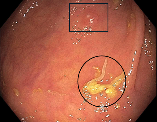
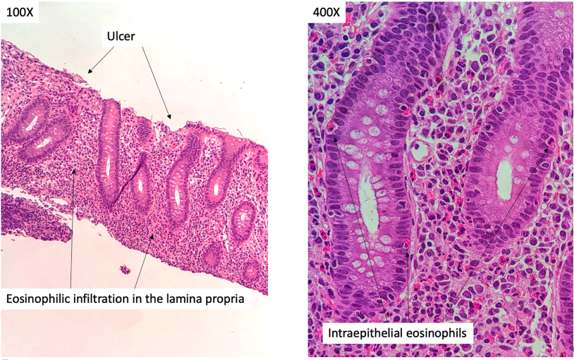
A Rare Cause of Gastrointestinal Hemorrhage: <i>Trichuris trichiura</i> Infection, ACG 2025 Annual Scientific Meeting Abstracts. Phoenix, AZ: American College of Gastroenterology.