Corewell Health William Beaumont University Hospital Royal Oak, MI
Samiksha Pandey, MD1, Ahmed Aref, MD1, Natalie Wilson, MD2, Fatima Elmustafa, MBBS3, Begum Akay, MD1, Mohamed Abdallah, MD1 1Corewell Health William Beaumont University Hospital, Royal Oak, MI; 2University of Minnesota, Minneapolis, MN; 3Henry Ford Warren, Warren, MI Introduction: Trichobezoars are rare gastrointestinal foreign bodies formed from ingested hair, typically occurring in adolescent females with psychiatric conditions such as trichophagia and trichotillomania. Endoscopic removal of trichobezoars is commonly unsuccessful and surgical management is required for most cases. Here, we present a case of a 10 cm trichobezoar that was found in a 15-year-old female.
Case Description/
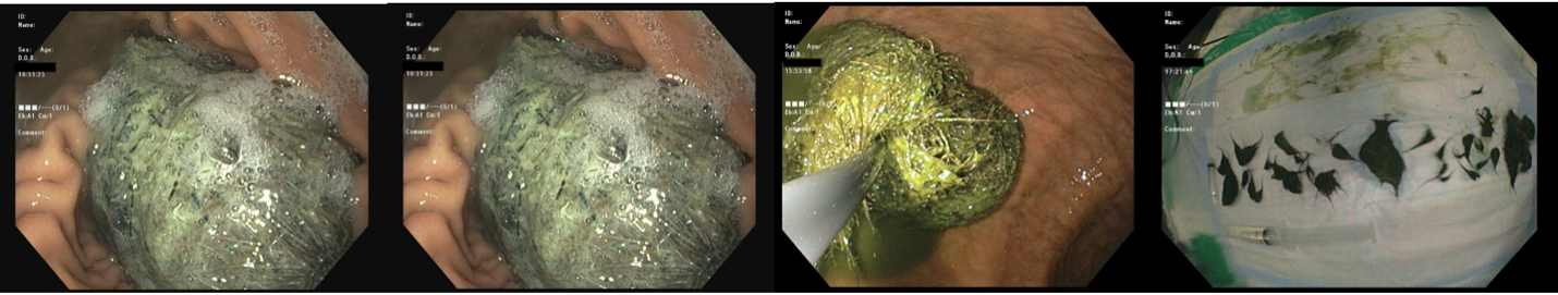
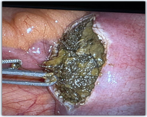
Methods: A 15-year-old female presented with abdominal pain, nausea, and vomiting. Computed tomography (CT) of the abdomen revealed retained gastric debris. An initial esophagogastroduodenoscopy (EGD) revealed a large, 10 cm gastric trichobezoar, of which only 20% was retrieved during a prolonged ( > 2 hours) endoscopic session using snares and forceps (Figure 1). The patient was instructed to consume carbonated beverages twice daily for two days to aid in bezoar dissolution. The patient underwent a repeat EGD two days later which lasted 2.5 hours and involved carbonated beverage injection into trichobezoar, and fragmentation with argon plasma coagulation (APC), in addition to mechanical fragmentation with snares and forceps (Figure 1). This resulted in complete endoscopic clearance of the gastric trichobezoar. The patient was discharged the following day, however, she presented again two days later to the ED with signs and symptoms of small bowel obstruction (SBO) confirmed with CT imaging showing a 9.4 cm trichobezoar lodged in the jejunum. The patientunderwent successful robotic-assisted laparoscopic enterotomy with bezoar extraction (Figure 2).Postoperative recovery was uneventful. The patient established care with the psychiatry team in the outpatient clinic. Discussion: This case underscores the therapeutic challenges of the management of trichobezoars. The use of chemical disintegration, coupled with APC fragmentation of trichobezoars with multiple endoscopic devices, even large trichobezoars, such as the 10 cm bezoar in our case, can be successfully removed endoscopically. This approach has been reported in the literature for management of similar cases of trichobezoars. However, our case demonstrates the potential adverse outcomes of this approach. Endoscopic removal of trichobezoar in one session using APC has been reported with excellent outcomes and may be more suitable for similar cases. A multidisciplinary approach, including medical, surgical, and psychiatric care, is essential for managing trichobezoar and preventing recurrence.
Figure: Figure 1: Trichobezoar in the stomach
Figure: Figure 2: Laparoscopic enterotomy with bezoar extraction
Disclosures: Samiksha Pandey indicated no relevant financial relationships. Ahmed Aref indicated no relevant financial relationships. Natalie Wilson indicated no relevant financial relationships. Fatima Elmustafa indicated no relevant financial relationships. Begum Akay indicated no relevant financial relationships. Mohamed Abdallah indicated no relevant financial relationships.
Samiksha Pandey, MD1, Ahmed Aref, MD1, Natalie Wilson, MD2, Fatima Elmustafa, MBBS3, Begum Akay, MD1, Mohamed Abdallah, MD1. P5170 - A Hairy Situation: Endoscopic Removal of Large Gastric Trichobezoar With a Jejunal Surprise, ACG 2025 Annual Scientific Meeting Abstracts. Phoenix, AZ: American College of Gastroenterology.

 photo")