Vassar Brothers Medical Center - Nuvance Health Poughkeepsie, NY
Bright Nwatamole, MBBS1, Muhammad Hassan, MD2, Simona Meca, MD3 1Vassar Brothers Medical Center - Nuvance Health, Poughkeepsie, NY; 2Nuvance Health, Poughkeepsie, NY; 3Premier Medical Group -GI Division, Poughkeepsie, NY Introduction: Foreign body ingestion is common among individuals with psychiatric illness; however, transmural impaction of sharp objects in the colon is exceedingly rare. We present a case of an asymptomatic, colonic wall–embedded sewing needle successfully retrieved via colonoscopy without the need for surgical intervention.
Case Description/
Methods: A 37-year-old incarcerated male with a history of bipolar disorder presented with vague abdominal discomfort and intermittent hematochezia two days following intentional ingestion of a sewing needle. He was hemodynamically stable, without signs of peritonitis. Physical examination, including rectal exam, and laboratory workup were unremarkable. Initial computed tomography (CT) of the abdomen revealed the sewing needle in the distal duodenum. Urgent esophagogastroduodenoscopy (EGD) with push enteroscopy was unable to localize the foreign body.
Serial abdominal radiographs over the subsequent five days demonstrated gradual migration of the needle through the small intestine, with final localization in the cecum. The needle remained in the same position for an additional five days. Repeat CT confirmed its presence within the cecal lumen.
On day 12 post-ingestion, colonoscopy was performed with surgical backup. After standard bowel preparation with polyethylene glycol, the needle was visualized embedded transmurally in a cecal mucosal fold opposite the ileocecal valve. The sharp end was lodged within the colonic wall, the shaft was piercing through the mucosa fold, while the eye of the needle protruded freely into the lumen. Using jumbo biopsy forceps, the needle was grasped by the eye, withdrawn fully into the colonoscope’s working channel, and removed without complication. No mucosal injury or bleeding was observed. The patient was monitored post-procedure and discharged in stable condition after 48 hours. Discussion: This case illustrates a rare presentation of a sharp foreign body embedded in the colonic wall without clinical signs of perforation. The successful endoscopic retrieval by enclosing the needle within the scope’s working channel highlights a safe and minimally traumatic technique. Timely imaging, multidisciplinary planning, and careful endoscopic intervention were critical in avoiding surgical management.
Figure: Colonoscopic view showing a sewing needle transmurally embedded in a cecal mucosal fold, with the eye of the needle protruding into the lumen and the sharp end lodged in the colonic wall.
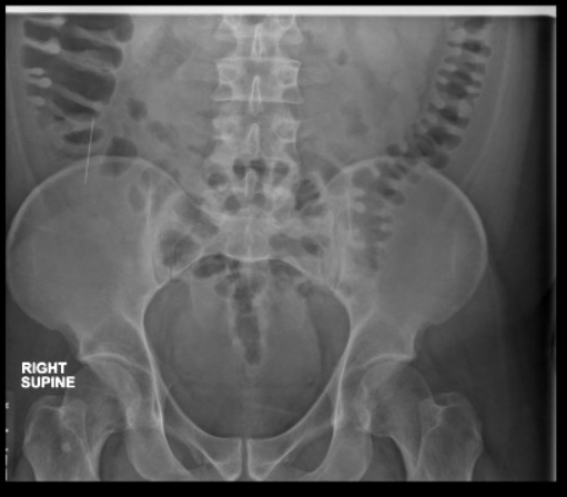
Figure: Supine Abdominal X-ray Demonstrating Radiopaque Linear Foreign Body (Sewing Needle) Localized to the Right Lower Quadrant
Disclosures: Bright Nwatamole indicated no relevant financial relationships. Muhammad Hassan indicated no relevant financial relationships. Simona Meca indicated no relevant financial relationships.
Bright Nwatamole, MBBS1, Muhammad Hassan, MD2, Simona Meca, MD3. P5148 - Silent Impactor: Colonoscopic Retrieval of a Cecal Wall-Embedded Sewing Needle in a Psychiatric Patient, ACG 2025 Annual Scientific Meeting Abstracts. Phoenix, AZ: American College of Gastroenterology.

 photo")