Mohammed Z. Rehman, MD1, Mariya Syed, MD1, Jayanthraj Gone, DO2, Shane Quo, DO2, Mohammed A. Ahmed, MBBS3, Faraz Eshaghi, DO2, Joseph Staffetti, MD2 1HCA Florida Healthcare, Land O Lakes, FL; 2HCA Florida Healthcare, Hudson, FL; 3Deccan College of Medical Sciences, Hyderabad, Telangana, India Introduction: Kommerell diverticulum is an extremely rare anomaly of an aberrant right or left subclavian artery in conjunction with a right or left-sided aortic arch, respectively, occurring in only about 0.7-2% of the population. This diverticulum is a remnant of the embryonic fourth aortic arch and is usually asymptomatic, but can cause a mass effect on the esophagus or trachea. Dysphagia lusoria is known as difficulty in swallowing when a vascular anomaly compresses the esophagus. We present a case of dysphagia lusoria, highlighting the presentation and diagnostic approach, and the importance of recognizing rare vascular anomalies as a cause of esophageal compression
Case Description/
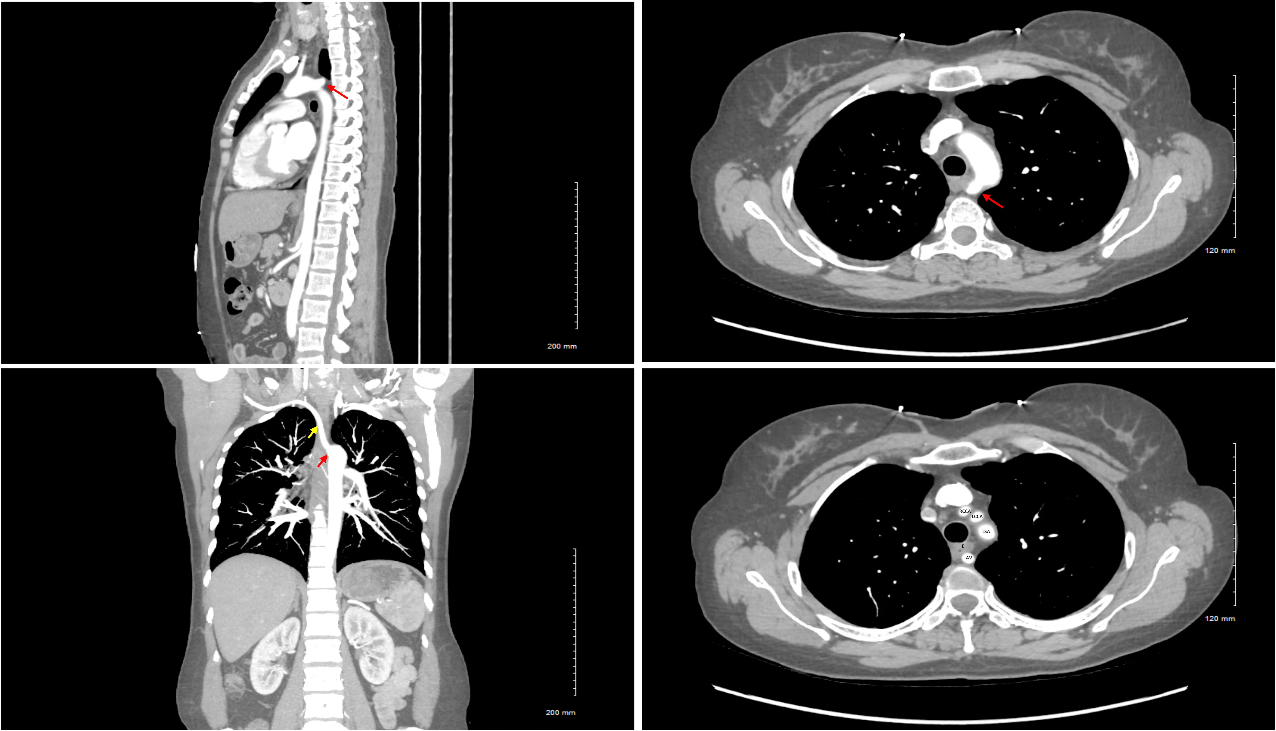
Methods: 40-year-old female with well-controlled seizure disorder, anxiety, smoking, and alcohol use admitted for dull chest pain, intermittent right upper quadrant pain, dizziness, and weakness. CT revealed an aberrant right subclavian artery with retroesophageal course and funneling of the origin compatible with Kommerell diverticulum (Image 1). On further questioning, the patient endorsed dysphagia to solids more than liquids, which has been present since “forever”, a foreign body sensation, and drooling saliva when she sleeps.
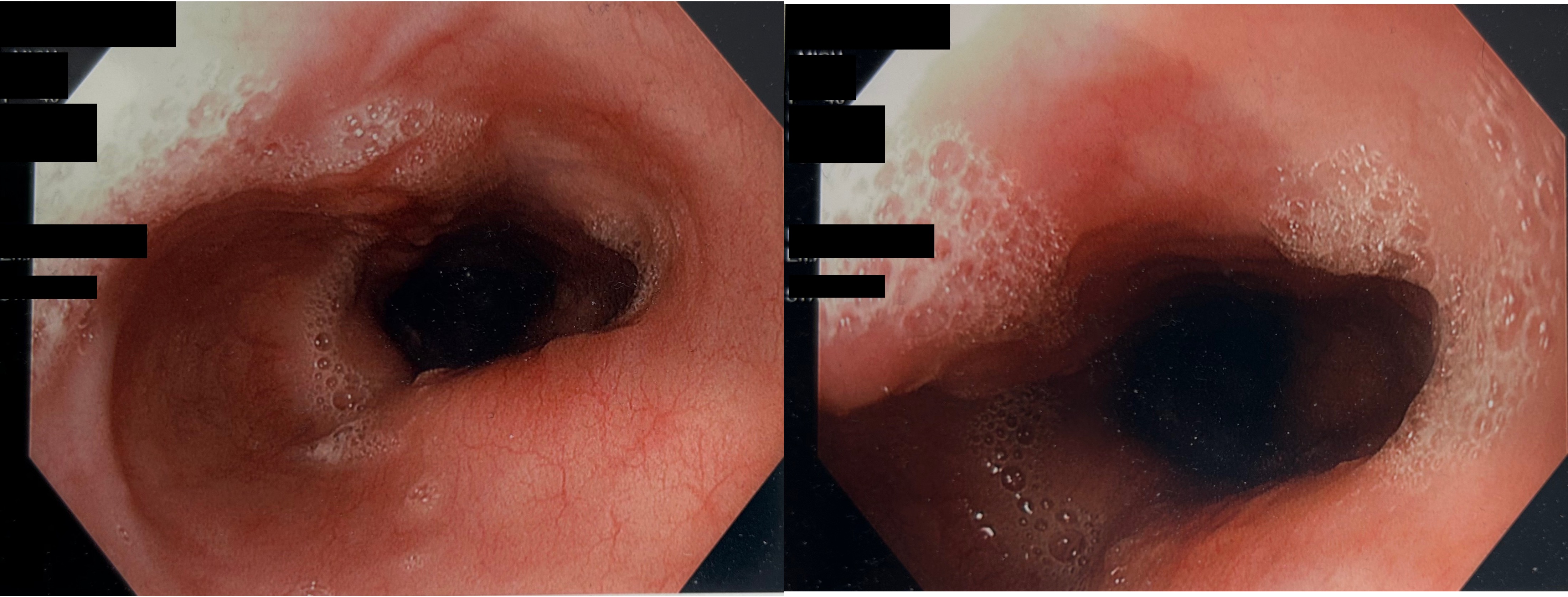
The patient underwent an esophagogastroduodenoscopy (EGD) to evaluate the dysphagia further, which revealed a pulsatile mass causing extrinsic compression at around 21 cm (Image 2). Given the relatively small size (less than 30mm), the patient was asked to make dietary and lifestyle modifications as surgical fixation was not recommended. Discussion: The extrinsic compression of the esophagus can lead to significant discomfort and progressive dysphagia. The patient’s symptoms of dysphagia were due to an aberrant right subclavian artery arising from a Kommerell diverticulum. This can be identified using CT or MRI imaging, while barium swallow and EGD are useful for evaluating the extent and nature of dysphagia. Treatment for asymptomatic or mildly symptomatic patients is controversial because of the rarity of the condition, and usually involves lifestyle modifications, such as in our case. Generally, surgical intervention is considered when the diameter of the diverticular orifice exceeds 30mm and/or the diameter of the descending aorta exceeds 50mm.
Kommerell diverticulum, though rare, should be recognized as a potential cause of chronic and unexplained dysphagia. This case emphasizes the importance of considering vascular anomalies in the differential diagnosis of dysphagia and tailoring management.
Figure: CT scan of the Chest with IV contrast Legend: Red arrow: Origin of the aberrant vessel, Kommerell’s diverticulum. Yellow Arrow: Course of the aberrant vessel. AV: Aberrant Vessel. E: Esophagus. LSA: Left Subclavian Artery. LCCA: Left Common Carotid Artery. RCCA: Right Common Carotid Artery.
Figure: EGD images
Disclosures: Mohammed Rehman indicated no relevant financial relationships. Mariya Syed indicated no relevant financial relationships. Jayanthraj Gone indicated no relevant financial relationships. Shane Quo indicated no relevant financial relationships. Mohammed Ahmed indicated no relevant financial relationships. Faraz Eshaghi indicated no relevant financial relationships. Joseph Staffetti indicated no relevant financial relationships.
Mohammed Z. Rehman, MD1, Mariya Syed, MD1, Jayanthraj Gone, DO2, Shane Quo, DO2, Mohammed A. Ahmed, MBBS3, Faraz Eshaghi, DO2, Joseph Staffetti, MD2. P5030 - Kommerell Diverticulum Causing Chronic Dysphagia: A Rare Vascular Anomaly, ACG 2025 Annual Scientific Meeting Abstracts. Phoenix, AZ: American College of Gastroenterology.