Corewell Health William Beaumont University Hospital Royal Oak, MI
Muhammad Ibrahim Majeed, MBBS1, Nishant Aggarwal, MD1, Fady Banno, MD1, Mohamed Abdallah, MD2, Mohammad Adam, MD, MSc3, Mehr Amer Majeed, MD4, Ali Osman, MD, MSCI Candidate5, Thuraya Mussa, MD6 1Corewell Health William Beaumont University Hospital, Royal Oak, MI; 2Corewell Health, Royal Oak, MI; 3University of Missouri - Kansas City School of Medicine, Kansas City, MO; 4SSM Health Saint Louis University Hospital, St. Louis, MO; 5Washington University School of Medicine in St. Louis, Ballwin, MO; 6Michigan State University, Lansing, MI Introduction: Transesophageal-echocardiography (TEE) is commonly used for detailed cardiac imaging of various cardiac conditions. While generally safe, it can occasionally lead to upper gastrointestinal (GI) adverse events. Here, we present a case of TEE-induced trauma that interestingly, presented as an esophageal mass lesion.
Case Description/
Methods: An 82-year-old female with a past medical history of paroxysmal atrial fibrillation on apixaban, coronary and valvular heart disease was evaluated for a three-day history of melena during a hospitalization for acute heart failure. She denied any dysphagia or prior history of GI bleeding. Her vitals were unremarkable. Physical examination showed pallor but she was otherwise well appearing. She had a TEE eight days prior to evaluation and her hemoglobin dropped from 8.3g/dL to 6.0g/dL in this timeframe. Esophagogastroduodenoscopy (EGD) one year prior had shown a hiatal hernia and erosive gastritis. Computed tomography (CT) chest, abdomen, pelvis revealed heterogeneity of sternum, thoracic and lumbar spine, suspicious for metastatic disease.
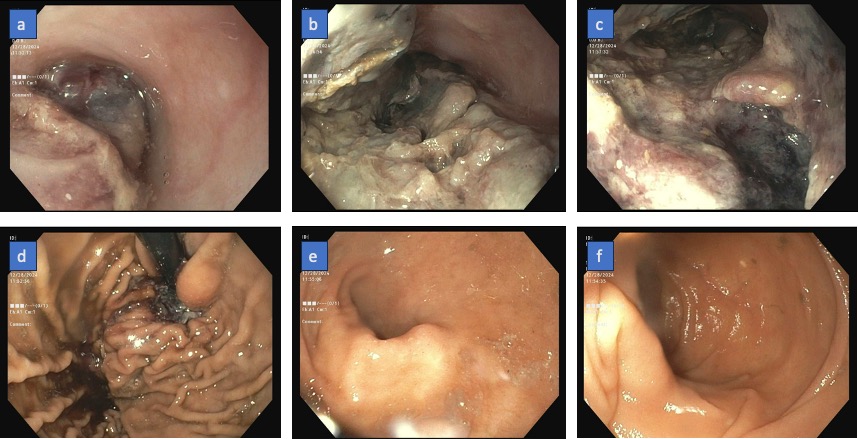
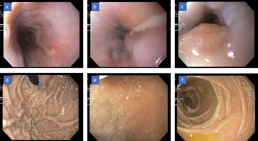
The patient received two units packed red blood cells, intravenous pantoprazole, and empiric octreotide; her apixaban was held. Due to suspicion of GI bleeding, EGD was performed and revealed a large, friable, ulcerated 13mm esophageal mass with necrotic debris and blood clots (figure 1). Small biopsies were obtained. Initial concern was traumatic ulceration of a possible underlying mass secondary to the recent TEE. Given no suspicion for varices, octreotide was discontinued. Biopsies revealed fragments of necrotic ulcer exudate with atypical squamous epithelium—deemed superficial and non-representative. Oncologic workup including SPEP, tumor markers, and nuclear bone scan were unremarkable. Her melena subsequently resolved, and she was managed conservatively with twice daily pantoprazole and close hemoglobin monitoring. Follow-up EGD two weeks later showed marked endoscopic improvement (figure 2), revealing only two superficial linear ulcers in the lower third of the esophagus, without evidence of mass or residual hematoma. Discussion: Though initially suggestive of malignancy, the esophageal mass was ultimately attributed to TEE-induced injury. To our literature review, we found 14 cases of TEE-related esophageal hematoma, emphasizing its rarity. Therefore, clinicians should be cautious with invasive instrumentation, particularly in elderly anticoagulated patients.
Figure: Figure 1: Initial EGD showing (a) friable ulcerated mass in the upper esophagus, (b) mid esophagus, (c) lower esophagus, (d) retroflexed view of gastric fundus, (e) pylorus, (f) second part of duodenum
Figure: Figure 2: Follow-up EGD two weeks after the initial exam showing (a) single linear clean based ulcer in the mid-esophagus, (b) ulcer extending to the lower esophagus, (c) gastroesophageal junction, (d) retroflexed view of the gastric fundus, (e) duodenal bulb, and (f) second part of duodenum
Disclosures: Muhammad Ibrahim Majeed indicated no relevant financial relationships. Nishant Aggarwal indicated no relevant financial relationships. Fady Banno indicated no relevant financial relationships. Mohamed Abdallah indicated no relevant financial relationships. Mohammad Adam indicated no relevant financial relationships. Mehr Amer Majeed indicated no relevant financial relationships. Ali Osman indicated no relevant financial relationships. Thuraya Mussa indicated no relevant financial relationships.
Muhammad Ibrahim Majeed, MBBS1, Nishant Aggarwal, MD1, Fady Banno, MD1, Mohamed Abdallah, MD2, Mohammad Adam, MD, MSc3, Mehr Amer Majeed, MD4, Ali Osman, MD, MSCI Candidate5, Thuraya Mussa, MD6. P5029 - Esophageal Mass on Upper Endoscopy Following Transesophageal Echocardiography, ACG 2025 Annual Scientific Meeting Abstracts. Phoenix, AZ: American College of Gastroenterology.

 photo")