Miguel Gomez, MD, Harmeet Malhi, MBBS, Douglas Simonetto, MD Mayo Clinic, Rochester, MN Introduction: Esophageal varices are a common complication of portal hypertension typically in the setting of liver cirrhosis. However, in the absence of portal hypertension, varices can occur due to mass effect and obstruction, with other rare causes including superior vena cava compression (SVC), mediastinal tumors, and thyroid masses.
Case Description/
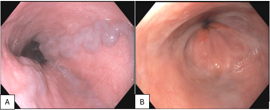
Methods: This is a 63-year-old female presenting to the hepatology clinic for isolated esophageal varices. For symptoms of worsening epigastric pressure and heartburn she had a prior diagnostic upper endoscopy (EGD) which demonstrated a large hiatal hernia and esophageal varices at an outside hospital. She had further undergone a transjugular liver biopsy and measurement of hepatic venous pressure gradient, both of which were reportedly normal. Liver biopsy slides were reviewed and reported normal lobular architecture with minimal steatosis and no fibrosis. Additionally, pathological inflammation, Mallory-Denk Bodies, and ballooning degeneration were absent in the liver biopsy. Trichrome stain was normal. Leading hypothesis on the cause of her isolated esophageal varices is SVC obstruction of unknown etiology or azygos vein compression from mass effect of her large hiatal hernia. EGD was repeated, and she was found to have two large columns of isolated distal esophageal varices (Figure 1A). SVC obstruction was ruled out with CT venogram, which showed a large hiatal hernia with extrinsic compression of the azygos vein. Patient underwent robotic assisted laparoscopic paraoesophageal hernia repair with partial fundoplication with thoracic surgery for symptom management. Clinic visit after surgery showed 50% reduction in reflux and pressure-like symptoms. Repeat EGD showed significantly decreased size of varices (Figure 1B). Discussion: Isolated esophageal varices are rare in the absence of portal hypertension. The most common cause is SVC compression from mediastinal or bronchogenic tumors resulting in the formation of “downhill” esophageal varices due to increased venous pressure. The azygous vein can undergo significant hemodynamic changes to drain varices. However, there are limited documented cases of azygous vein compression leading to esophageal varices.
Figure: Figure 1: A) EGD before paraoesophageal hernia repair B) EGD after paraoesophageal hernia repair
Disclosures: Miguel Gomez indicated no relevant financial relationships. Harmeet Malhi indicated no relevant financial relationships. Douglas Simonetto indicated no relevant financial relationships.
Miguel Gomez, MD, Harmeet Malhi, MBBS, Douglas Simonetto, MD. P5023 - Downhill Danger: A Rare Case of Esophageal Varices From Hiatal Hernia-Induced Compression, ACG 2025 Annual Scientific Meeting Abstracts. Phoenix, AZ: American College of Gastroenterology.


 photo")