California Pacific Medical Center San Francisco, CA
Yohanna Khachiyan, MD1, Matthew Bell, MD1, Benjamin Yip, MD2 1California Pacific Medical Center, San Francisco, CA; 2Sutter Health, San Francisco, CA Introduction: Herpes Simplex virus (HSV) esophagitis is a well recognized viral infection in immunocompromised hosts. The most common findings on upper endoscopy are multiple, well-circumscribed, shallow ulcers with a “volcano-like” appearance. We present a case of HSV esophagitis in an immunocompromised host with unusual findings on upper endoscopy.
Case Description/
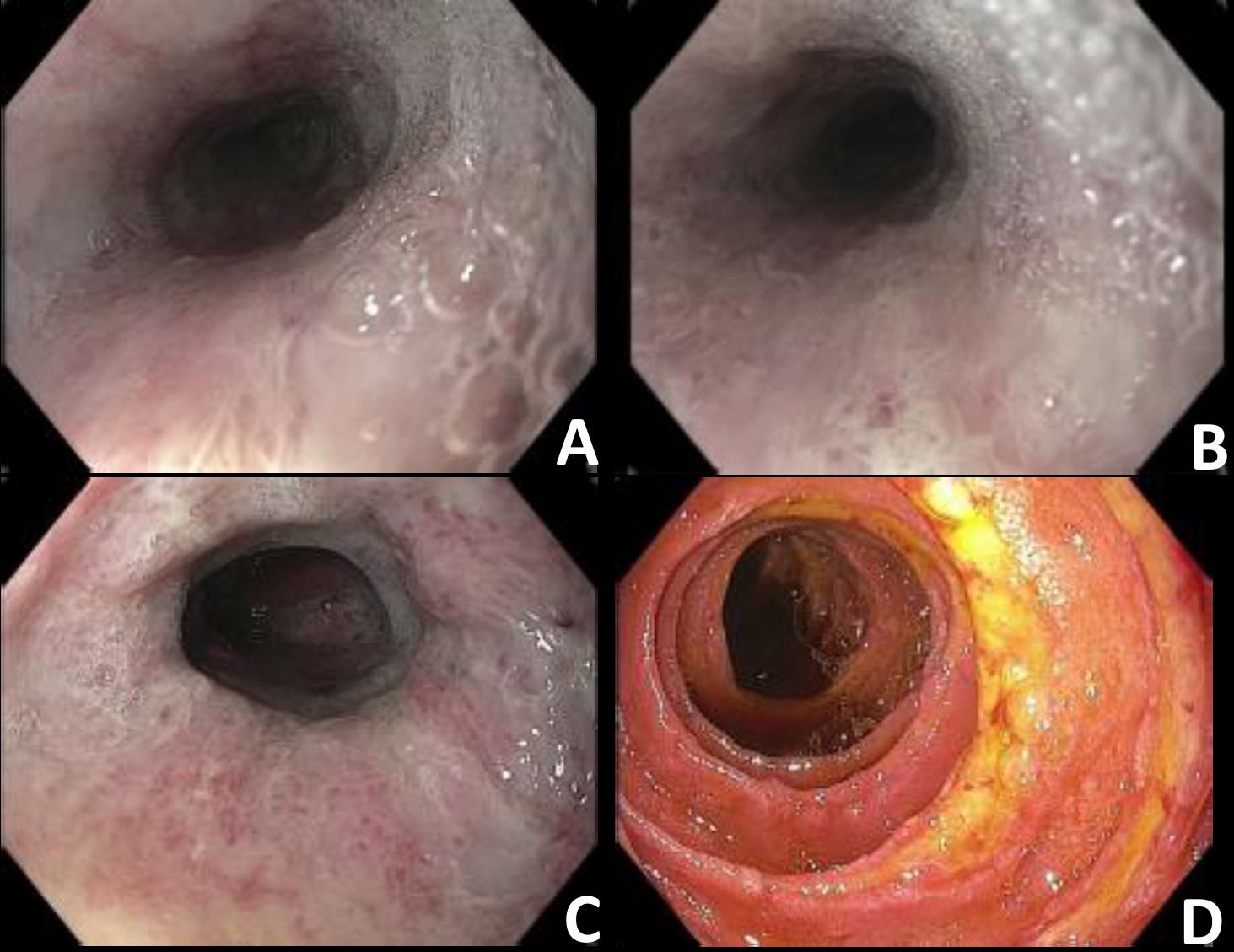
Methods: A 66-year-old male patient with a past medical history of ANCA-positive vasculitis complicated by end-stage renal disease status post kidney transplant (on tacrolimus and mycophenolate mofetil) presented with abdominal pain, odynophagia, nausea, and vomiting. Upper endoscopy from an outside hospital showed a diffusely ulcerated esophagus with scattered white plaques and a black mucous layer coating the entire esophagus as well as multiple duodenal ulcers with a black pigmented base. Biopsies showed ulcerated stromal tissue with necrosis and severe acute inflammation. CMV/HSV staining was not done. The patient was treated with fluconazole, pantoprazole 40 mg twice daily, and methylprednisolone IV for suspected ANCA vasculitis. He was ultimately transferred to our hospital due to a lack of improvement in symptoms. Repeat endoscopy showed severe, diffuse, pan-esophageal esophagitis and friability with mucosal sloughing (Figure 1: A, B, C) along with clean based (Forest III) linear duodenal ulcers (Figure 1: D). Biopsies with immunohistochemical staining were positive for HSV. He was started on IV acyclovir with improvement in his symptoms and he was discharged with oral valacyclovir. Discussion: HSV esophagitis is a well-known condition affecting immunocompromised individuals. Many case reports and series describe its typical endoscopic findings of superficial ulcers most often involving the distal esophagus. Although this patient had a classic clinical presentation for infectious esophagitis, his endoscopic findings of severe pan-esophageal esophagitis were atypical for HSV. It is important to have a high suspicion for infectious esophagitis in any immunocompromised patient who presents with odynophagia and to request viral staining despite having an atypical endoscopic appearance.
Figure: Esophageal findings from upper endoscopy.
Disclosures: Yohanna Khachiyan indicated no relevant financial relationships. Matthew Bell indicated no relevant financial relationships. Benjamin Yip indicated no relevant financial relationships.
Yohanna Khachiyan, MD1, Matthew Bell, MD1, Benjamin Yip, MD2. P5005 - An Unusual Presentation of Pan-Esophageal Esophagitis Due to Herpes Simplex Virus in an Immunocompromised Patient, ACG 2025 Annual Scientific Meeting Abstracts. Phoenix, AZ: American College of Gastroenterology.

 photo")