Rahul Patel, DO, Sandus Khan, MD Northeast Georgia Medical Center, Gainesville, GA Introduction: Hypopharyngeal cancers are relatively rare cancers, with only a few thousand cases diagnosed each year in the U.S. Presenting symptoms are often vague, such as dysphagia, odynophagia, weight loss, bleeding. Squamous cell carcinomas account for 95% of cases and prognosis is generally poor as most are diagnosed at late-stage. Hypopharyneal cancers are already difficult to diagnose and the more common diagnoses divert clinical attention away, leading to delayed or missed diagnosis. We present such a case where a hypopharyngeal mass was initially missed due to a coexisting diagnosis of esophageal varices.
Case Description/
Methods: A 61 yo female with past medical history significant for alcoholic liver cirrhosis with esophageal varices, history of C. difficile colitis presented to ED with acute onset hematemesis and worsening abdominal pain for past few hours. Patient required ICU admit as she was hypotensive, continued hematemesis, and severe anemia. EGD was performed and showed 3 columns of small varies with overlying red wale markings but no obvious active blood noted. 2 bands were placed for varices and while withdrawing the scope there was persistent blood noted in the hypopharyngeal region, no further investigation performed at that time. Patient initially improved, but then had continued hematemesis and hypotension following day requiring repeat EGD. Repeat EGD showed excess blood in hypopharyngeal region, which now prompted significant suctioning for further evaluation and a polypoid mass lesion in the right piriform fossa with active oozing was noted. ENT was consulted and direct laryngoscopy with biopsy performed and showed invasive squamous cell carcinoma, focally keratinizing, p16 negative. Heme/onc was consulted and reported unable to start chemotherapy or radiation therapy given other comorbidities. Surgical evaluation for possible resection was offered but given overall poor prognosis patient and family decided on pursing hospice care. Discussion: Hypopharyngeal carcinomas are rare but aggressive malignancies that are often diagnosed at a late stage given vague and insidious onset of symptoms. However, in this specific case the diagnosis was further delayed due to coexisting cirrhosis with esophageal varices thought to be the culprit of hematemesis. Patient had presented with similar symptoms prior to this hospital visit and possibly with more vigilance the hypopharyngeal mass could have been detected earlier with better outcomes for the patient.
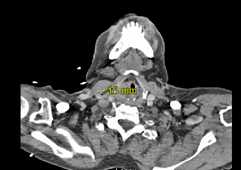
Figure: CT neck showing hypopharyngeal mass within the right piriform sinus
Disclosures: Rahul Patel indicated no relevant financial relationships. Sandus Khan indicated no relevant financial relationships.
Rahul Patel, DO, Sandus Khan, MD. P4999 - A Diagnostic Overshadowing: Hypopharyngeal Malignancy Masked by Esophageal Varices, ACG 2025 Annual Scientific Meeting Abstracts. Phoenix, AZ: American College of Gastroenterology.

.jpg "Rahul Patel, DO (he/him/his) photo")