
Tuesday Poster Session
Category: Colon

Mustafa Naveed, DO
Virginia Tech Carilion School of Medicine
Roanoke, VA
Rectal squamous cell carcinoma (SCC) is a rare epithelial malignancy accounting for 0.3% of rectal cancers, with less than 150 cases reported. It predominantly affects older adults and is slightly more common in females. The 5-year survival rate is 32%, varying by stage. Endoscopically, rectal SCC often appears as a friable, ulcerated, or exophytic lesion near the dentate line. We present a case of rectal SCC with an unusual vascular, eroded appearance highlighting the importance of recognizing atypical presentations.
Case Description/
Methods:
A 77-year-old female with a history of diverticulosis, constipation presented with lightheadedness, fatigue, and intermittent bright red blood per rectum. Objective data demonstrated a hemoglobin drop from 13.7 g/dL to 10.9 g/dL. Her previous colonoscopy, done three years prior, was notable only for four benign colonic polyps being removed. Recommendation at that time was for a repeat colonoscopy in three years. Surveillance colonoscopy revealed mild sigmoid diverticulosis and an anorectal lesion with a vascular, eroded surface near the dentate line (Figure 1). Biopsies were taken, and local bleeding was controlled with a hemoclip. Pathology showed high-grade squamous intraepithelial lesion (AIN 3), positive for p40, p16, and p53-null pattern. She was discharged in stable condition with colorectal surgery follow-up.
Discussion:
This case highlights the diagnostic challenges of an anorectal lesion with an unusual vascular and eroded appearance (Figure 1), initially raising concern for benign or malignant processes. The differential diagnosis included hemorrhoids, anal fissures, inflammatory polyps, and rectal SCC. Rectal SCC is rare and often presents as an ulcerated or exophytic mass, but in this case its vascular and eroded appearance was atypical. Pathology confirmed AIN 3, consistent with SCC precursor. Definitive diagnosis requires biopsy, but caution should be taken when biopsying these lesions due to their propensity to bleed given their vascular nature. Hemostatic options typically allow for safe biopsy and bleeding control. Treatment can be curative in intent and includes options such as: local excision, low anterior resection, and/or abdominoperineal resection with chemoradiation therapy. This case aims to familiarize endoscopists with a unique presentation of rectal SCC, remind endoscopists to maintain a broad differential when evaluating lesions near the dentate line, and emphasize early recognition to avoid delays in appropriate management. 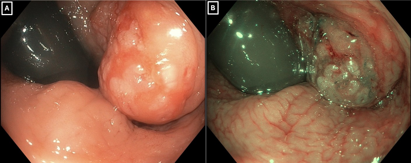
Figure: Figure 1: A & B) Endoscopic image demonstrating retroflexed view of an anorectal lesion with a vascular, eroded surface near the dentate line
Disclosures:
Mustafa Naveed indicated no relevant financial relationships.
Waleed Iftikhar indicated no relevant financial relationships.
Jonathan Rozenberg indicated no relevant financial relationships.
Subhash Garikipati indicated no relevant financial relationships.
Bara El Kurdi indicated no relevant financial relationships.
Mustafa Naveed, DO1, Waleed Iftikhar, BS2, Jonathan Rozenberg, DO1, Subhash Garikipati, MD3, Bara El Kurdi, MD3. P4738 - An Atypical Presentation of Rectal Squamous Cell Carcinoma, ACG 2025 Annual Scientific Meeting Abstracts. Phoenix, AZ: American College of Gastroenterology.