Georgetown University MedStar Health Baltimore, WA
Shahem Abbarh, MD1, Bisher Sawaf, MD2, Ashraf Ahmed, MD3, Obada Daaboul, MD4, Mhd Kutaiba Albuni, MD5, Yusuf Omar Hallak, MD6, Muhamad Hijazi, MD7, Muaz Alsabbagh, MD8, Omar Brijawi, MD9, Amine Rakab, MD10 1Georgetown University MedStar Health, Baltimore, WA; 2University of Toledo Medical Center, Toledo, OH; 3College of Medicine, QU Health. Qatar University, Doha, Qatar., Doha, Ad Dawhah, Qatar; 4Southern Illinois University, Springfield, IL; 5Department of Internal Medicine, TriHealth Inc., Cincinnati, Cincinnati, OH; 6The University of Toledo, Toledo, OH; 7TriHealth, Cincinnati, OH; 8Detroit Medical Center/Wayne State University, Cleveland, OH; 9Mount Carmel Health Systems, Grove City, OH; 10Division of Medical Education, Weill Cornell Medicine, Doha, Ad Dawhah, Qatar Introduction: Hepatocellular carcinoma (HCC) with right atrial (RA) involvement is a rare but aggressive presentation, often associated with poor prognosis. Standardized treatment guidelines for such cases are lacking due to their rarity.
Case Description/
Methods: We report the case of a 68-year-old male with a history of hepatitis C-related cirrhosis who was incidentally found to have a right atrial mass during a preoperative evaluation. Imaging and histopathological studies confirmed the diagnosis of metastatic HCC. The patient underwent partial surgical resection of the mass, followed by adjuvant immunotherapy with atezolizumab and bevacizumab. Upon disease progression, he received targeted radiotherapy and was subsequently started on oral lenvatinib. Follow-up imaging showed marked regression of the cardiac mass and no evidence of intrahepatic recurrence. Discussion: This case highlights the value of a multidisciplinary, multimodal treatment approach, including surgery, immunotherapy, radiotherapy, and targeted therapy, for managing advanced HCC with cardiac involvement. It also underscores the need for heightened clinical suspicion and individualized therapeutic strategies in cases with atypical presentations
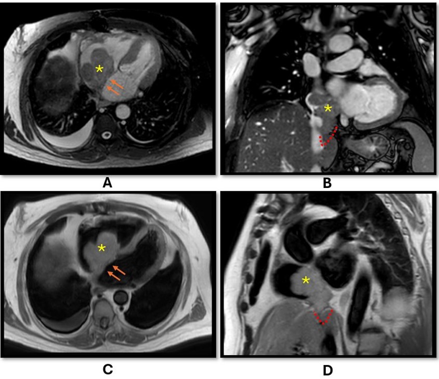
Figure: A and B: Axial and Coronal Balanced Turbo Field Echo (BTFE), respectively. C: Black Blood Single Shot Breath Hold (BB-SSh-BH). D: T2 Black Blood Short Axis Single Shot (T2-BB-SA-SSh). The images show large lobulated mass occupying the right atrium (Asterisk) attached to the interatrial septum (Arrows) with inferiorly extending component to the suprahepatic IVC (Dashed Red). The largest dimension of the lesion (craniocaudal) was approximately 6.8 cm.
Disclosures: Shahem Abbarh indicated no relevant financial relationships. Bisher Sawaf indicated no relevant financial relationships. Ashraf Ahmed indicated no relevant financial relationships. Obada Daaboul indicated no relevant financial relationships. Mhd Kutaiba Albuni indicated no relevant financial relationships. Yusuf Omar Hallak indicated no relevant financial relationships. Muhamad Hijazi indicated no relevant financial relationships. Muaz Alsabbagh indicated no relevant financial relationships. Omar Brijawi indicated no relevant financial relationships. Amine Rakab indicated no relevant financial relationships.
Shahem Abbarh, MD1, Bisher Sawaf, MD2, Ashraf Ahmed, MD3, Obada Daaboul, MD4, Mhd Kutaiba Albuni, MD5, Yusuf Omar Hallak, MD6, Muhamad Hijazi, MD7, Muaz Alsabbagh, MD8, Omar Brijawi, MD9, Amine Rakab, MD10. P5974 - A Rare Case of Hepatocellular Carcinoma Presenting With Right Atrial Involvement: Clinical Challenges and Multimodal Treatment, ACG 2025 Annual Scientific Meeting Abstracts. Phoenix, AZ: American College of Gastroenterology.

 photo")