Lorimar Rodriguez, MD1, Gretchen C.. Abarca-Riancho, MD2, Reinaldo Ramirez, MD1, Carlos Rosario, MD1 1Damas Hospital, Ponce, Puerto Rico; 2Damas Hospital, Guaynabo, Puerto Rico Introduction: Gastric xanthomas (GX) are benign mucosal lesions characterized by accumulation of foamy macrophages within the lamina propria of the gastrointestinal tract. While their clinical significance remains unclear, they have been associated with chronic gastritis, Helicobacter pylori infection, gastric atrophy, and gastric malignancy. GX are rare, with an incidence of 0.02-0.8% as incidental finding on endoscopy. Although usually benign, their presence may prompt concern for serious pathology. These lesions may also predispose patients to gastric malignancy, though their role to predict malignancy is unknown.
Case Description/
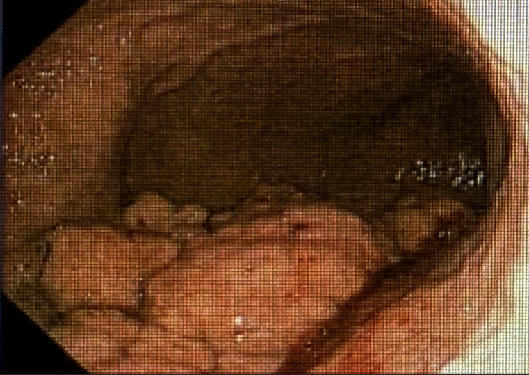
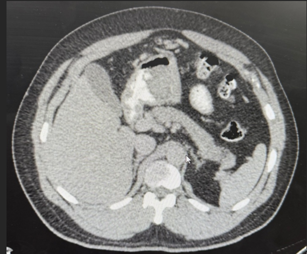
Methods: We present a 44 years old male Puerto Rican patient with past medical history of chronic gastritis who presents to the ED complaining of left lower quadrant pain. He denied weight loss, hematochezia or melena. Physical examination was remarkable for LLQ tenderness without peritoneal signs. Laboratory findings included leukocytosis (13.8) with neutrophilic predominance and elevated C-reactive protein. Electrolytes, urinalysis, liver enzymes, amylase, and lipase were within normal parameters. Abdominal CT scan (Fig. 1) showed findings consistent with sigmoid diverticulitis and a large soft tissue mass in the gastric body. The patient was admitted and treated with IV antibiotics and scheduled for esophagogastroduodenoscopy. EGD showed a large lesion on the posterior wall of the stomach. Histologic examination (Fig.2) was consistent with gastric xanthoma and H. pylori. The patient was discharged with treatment prescription and appointment with gastroenterology. Discussion: GX are non-neoplastic lesions mostly found on the stomach in patients with underlying mucosal pathology. Although often asymptomatic and incidental, their clinical relevance arises from their association with gastric diseases such as H pylori, chronic gastritis, and gastric malignancy. This patient's history of H pylori and gastritis is consistent with risk factors for GX. These lesions are often misidentified as malignancies during EGD due to their appearance and should prompt further evaluation with biopsy or imaging. The incidental finding of GX demonstrates the importance of considering benign etiologies in the differential diagnosis, especially in patients with known risk factors. While treatment is not typically needed, GX may mimic serious pathology and should be further investigated. Clinicians should be aware of the association between GX, H pylori and malignancy when evaluating similar cases.
Figure: Figure 1. Abdominal CT scan: large soft tissue gastric mass
Figure: Figure 2. EGD: large irregular mass on posterior wall of the stomach
Disclosures: Lorimar Rodriguez indicated no relevant financial relationships. Gretchen Abarca-Riancho indicated no relevant financial relationships. Reinaldo Ramirez indicated no relevant financial relationships. Carlos Rosario indicated no relevant financial relationships.
Lorimar Rodriguez, MD1, Gretchen C.. Abarca-Riancho, MD2, Reinaldo Ramirez, MD1, Carlos Rosario, MD1. P6417 - Incidental Detection of Gastric Xanthoma in a Middle-Aged Puerto Rican Patient With Acute Diverticulitis, ACG 2025 Annual Scientific Meeting Abstracts. Phoenix, AZ: American College of Gastroenterology.