University of Arizona College of Medicine Tucson, AZ
Samuel Cheong, DO1, Maxwell Hart, MD2, Karen Rico, MD1, Natasha Khona, DO1, Avin Aggarwal, MD1 1Banner - University of Arizona Tucson, Tucson, AZ; 2University of Arizona College of Medicine, Tucson, AZ Introduction: Malignant melanoma is the most aggressive form of skin cancer, characterized by high metastatic potential to any organ; however, metastasis to the gastrointestinal (GI) tract is rare, particularly to the stomach. We present a rare case of an 82-year-old male with a history of melanoma who was later found to have metastatic gastric melanoma.
Case Description/
Methods: An 82-year-old male with a history of melanoma of the left upper arm (stage IIIB) had undergone wide local excision with sentinel lymph node biopsy revealing two positive nodes and had been on pembrolizumab therapy for one year. He presented with a 2-week history of fatigue. Initial workup showed hemoglobin of 8.8 g/dL and MCV of 98 fL. Iron panel revealed iron of 32 μg/dL, transferrin saturation of 8%, and ferritin of 18 ng/mL, consistent with iron deficiency anemia (IDA). He denied abdominal pain, nausea, vomiting, melena, or hematochezia.
Initial EGD and colonoscopy were performed for workup of IDA without evidence of overt gastrointestinal bleeding. EGD revealed several 3–5 mm mucosal papules (nodules) with central pigmentation and stigmata of bleeding in the gastric fundus and body. Biopsies demonstrated brown-black pigmented neoplastic cells with immunohistochemical staining positive for SOX10 and Melan-A, consistent with metastatic melanoma. A subsequent colonoscopy was unremarkable.
One month later, the patient was admitted with new-onset melena. Repeat EGD showed four small to medium-sized, polypoid, non-circumferential masses with oozing and stigmata of recent bleeding on the lesser curvature of the stomach. Hemostatic spray and clips were used to control the bleeding. The patient was started on omeprazole 40 mg twice daily indefinitely, and radiation oncology was consulted for further management. Discussion: Metastatic gastric melanoma is a rare manifestation, accounting for only 2–4% of all melanoma metastases, with the stomach less commonly involved than the small intestine. Patients are usually asymptomatic at presentation. Endoscopic findings typically reveal pigmented or amelanotic polypoid lesions, and diagnosis is confirmed through histopathology and immunohistochemical staining, including markers such as S100, HMB-45, SOX10, and Melan-A. Prognosis remains poor, with median survival of 4 to 6 months and 5-year survival rates below 10%. Timely diagnosis with EGD and colonoscopy is essential for guiding treatment strategies, improve quality of life by mitigating complications, and increase survival in these patients.
Figure: Figure 1: A, B, C. A few 3 to 5 mm mucosal papules with central pigmentation found in the gastric fundus and gastric body demonstrating gastric melanoma. D. Medium-sized polypoid, non-circumferential mass with oozing bleeding consistent with gastric melanoma found on the lesser curvature of the stomach.
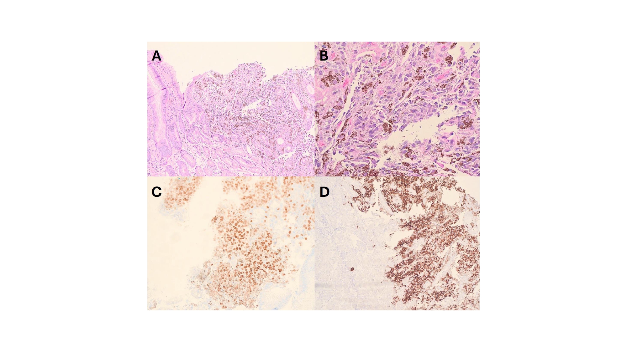
Figure: Figure 2: A. Gastric biopsy shows benign gastric mucosa adjacent to an area of ulceration with an infiltrate of malignant cells and pigment-laden macrophages. [H&E x10] B. The malignant cells are pleomorphic, hyperchromatic and occasionally have prominent nuclei. [H&E x40] C. Sox10 immunohistochemical stains show positive nuclear staining in the malignant cells. [H&E x20] D. Melan A immunohistochemical stains shows positive cytoplasmic staining in the malignant cells. [H&E x10]
Disclosures: Samuel Cheong indicated no relevant financial relationships. Maxwell Hart indicated no relevant financial relationships. Karen Rico indicated no relevant financial relationships. Natasha Khona indicated no relevant financial relationships. Avin Aggarwal indicated no relevant financial relationships.
Samuel Cheong, DO1, Maxwell Hart, MD2, Karen Rico, MD1, Natasha Khona, DO1, Avin Aggarwal, MD1. P6366 - Black Mystery in the Belly: When Melanoma Strikes the Stomach, ACG 2025 Annual Scientific Meeting Abstracts. Phoenix, AZ: American College of Gastroenterology.