University of Arizona College of Medicine Tucson, AZ
Celeste R. Gracey, DO1, Samuel Cheong, DO2, William Matloff, MD, PhD2, Franklin Liu, MD1, Sabrina Ho, MD1, Brandon Witten, MD2 1University of Arizona College of Medicine, Tucson, AZ; 2Banner - University of Arizona Tucson, Tucson, AZ Introduction: Primary appendiceal tumors are rare, with an incidence of 0.12 cases per 1,000,000 annually. Histological subtype is an important prognostic factor, with signet-ring cell carcinoma associated with a 27% 5-year survival rate. These tumors are typically unsuspected until appendectomy. We present an unusual case of appendiceal adenocarcinoma with signet-ring features, initially identified in the biliary system.
Case Description/
Methods: A 61-year-old woman presented with fatigue, low appetite, and weight loss. Computed tomography scan showed a hilar mass encasing the bile duct and hepatic vasculature, with peritoneal and ovarian metastases. ERCP with stenting was performed, but biliary brushings were negative. Ultrasound-guided liver biopsy revealed adenocarcinoma with signet-ring features, CK20 positive and focally CK7 positive, suggesting a lower gastrointestinal origin. Colonoscopy revealed a 3 cm, fungating cecal mass; biopsies matched the hepatic metastases. Palliative chemotherapy was initiated, and the patient was managed with biliary stenting for 18 months before transitioning to hospice care. Discussion: Appendiceal cancers are rare and often diagnosed incidentally after surgery for appendicitis. Our case is notable for its initial presentation as biliary obstruction, a pattern typically seen with cholangiocarcinoma or pancreatic cancer. The signet-ring cell subtype, known for aggressive behavior and diffuse peritoneal spread, initially mimicked gynecologic malignancies, further complicating diagnosis. Biliary system metastasis from an appendiceal primary is extremely rare. This highlights the importance of immunohistochemistry in establishing the tumor’s origin. CK20 positivity, focal CK7 expression, and patchy neuroendocrine marker staining provided crucial diagnostic clues linking the hepatic metastasis to an appendiceal source. This case underscores the diagnostic challenges when appendiceal tumors present with atypical features. In patients with biliary obstruction and metastatic adenocarcinoma of unknown primary, lower gastrointestinal sources should be considered. Early recognition is vital for guiding appropriate treatment and avoiding misdirected therapies. Given the rarity of such cases, further study is warranted to clarify metastatic pathways and improve diagnostic strategies for appendiceal cancers.
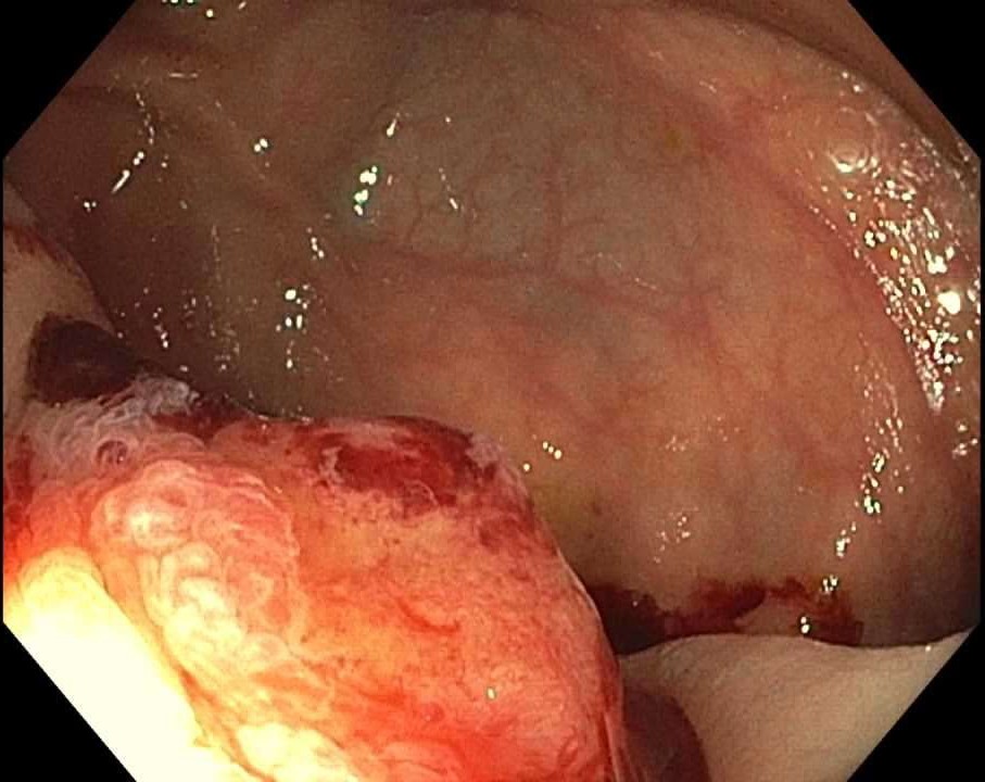
Figure: Colonoscopy identified a 3 cm, fungating cecal mass. A biopsy was found to have CK20 positivity, focal CK7 expression, and patchy neuroendocrine marker, consistent with a primary appendiceal tumor.
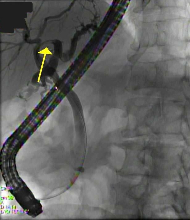
Figure: Imaging from ERCP demonstrates a non-obstructing biliary stricture of the left main hepatic duct in a patient with primary appendiceal cancer metastasized to the biliary system.
Disclosures: Celeste Gracey indicated no relevant financial relationships. Samuel Cheong indicated no relevant financial relationships. William Matloff indicated no relevant financial relationships. Franklin Liu indicated no relevant financial relationships. Sabrina Ho indicated no relevant financial relationships. Brandon Witten indicated no relevant financial relationships.
Celeste R. Gracey, DO1, Samuel Cheong, DO2, William Matloff, MD, PhD2, Franklin Liu, MD1, Sabrina Ho, MD1, Brandon Witten, MD2. P2578 - A Hidden Culprit: Biliary Metastasis Revealing an Appendiceal Signet-Ring Cell Carcinoma, ACG 2025 Annual Scientific Meeting Abstracts. Phoenix, AZ: American College of Gastroenterology.