
Monday Poster Session
Category: Colon
.jpg "Mohammed S. Zaman, DO photo")
Mohammed S. Zaman, DO
Mercyhealth Rockford
Rockford, IL
Colonic neuroendocrine carcinoma (NEC) is a rare, aggressive malignancy that usually presents in advanced stages, featuring rapid growth, local invasion, and distant metastasis. Median overall survival is typically less than nine months. We report a case of poorly differentiated primary colonic NEC with small cell features.
Case Description/
Methods:
A 49-year-old male with history of tobacco use presented after three days of worsening, diffuse abdominal pain and constipation. In the emergency department, vitals were stable, but WBC was 23.3K U/L. CT abdomen/pelvis with IV contrast revealed a large left upper quadrant mass (23.7 x 14.4 x 11.3 cm) with central necrosis, ill-defined hepatic lesions, a left lung base mass (4.3 x 1.7 x 3 cm), and significant lymphadenopathy.
Endoscopic evaluation via EGD demonstrated severe esophagitis and gastritis. Colonoscopy identified a 7 cm fungating, infiltrative mass at the splenic flexure causing partial luminal obstruction and oozing. Biopsies of both colonic and hepatic lesions confirmed high-grade, poorly differentiated NEC. Immunohistochemistry was positive for synaptophysin and CD56 with a Ki-67 index >95%, but negative for chromogranin, CDX2, and CK20.
Clinical course was complicated by sepsis and intra-abdominal abscess with colonic fistulization, requiring percutaneous drain placement. Repeat CT showed expansion of the mass to 32 x 18 x 21 cm. Despite broad-spectrum IV antibiotics and plans for carboplatin/etoposide chemotherapy, the extent of metastatic disease precluded surgical intervention, prompting early goals-of care discussions and transition to hospice.
Discussion:
Colonic NECs account for < 1% of colorectal cancers. Due to their aggressive growth pattern and proclivity for both infiltration as well as metastasis, multiple peritoneal structures may become involved concomitantly. This often necessitates advanced endoscopic techniques for visualization and mitigation of intestinal complications such as obstruction. The massive tumor size at diagnosis ( > 30 cm) alongside metastatic disease and rare complications of both fistula and abscess formation highlight this report as an exceptionally severe case. Although platinum-based chemotherapy offers modest response rates (~40–50%), outcomes remain poor, with median survival under 12 months. This case highlights the challenges in addressing advanced colonic NEC and the paramountcy of early, multidisciplinary intervention to improve management strategies.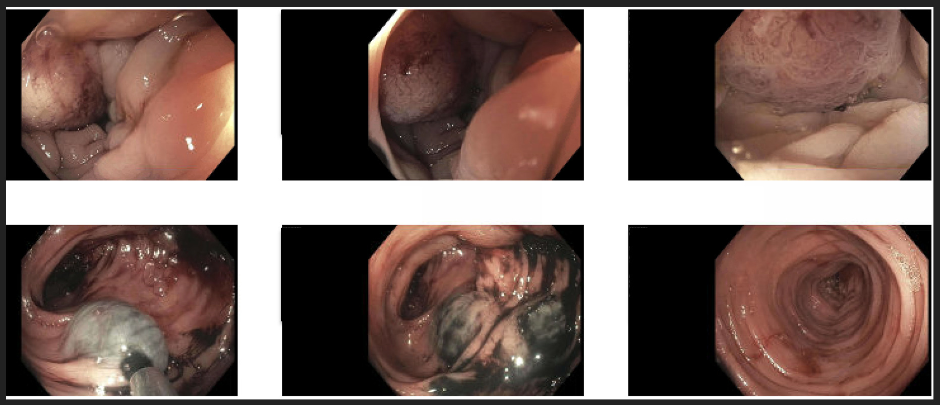
Figure: CT abdomen/pelvis imaging of large left upper quadrant mass (23.7 x 14.4 x 11.3 cm) with central necrosis, ill-defined hepatic lesions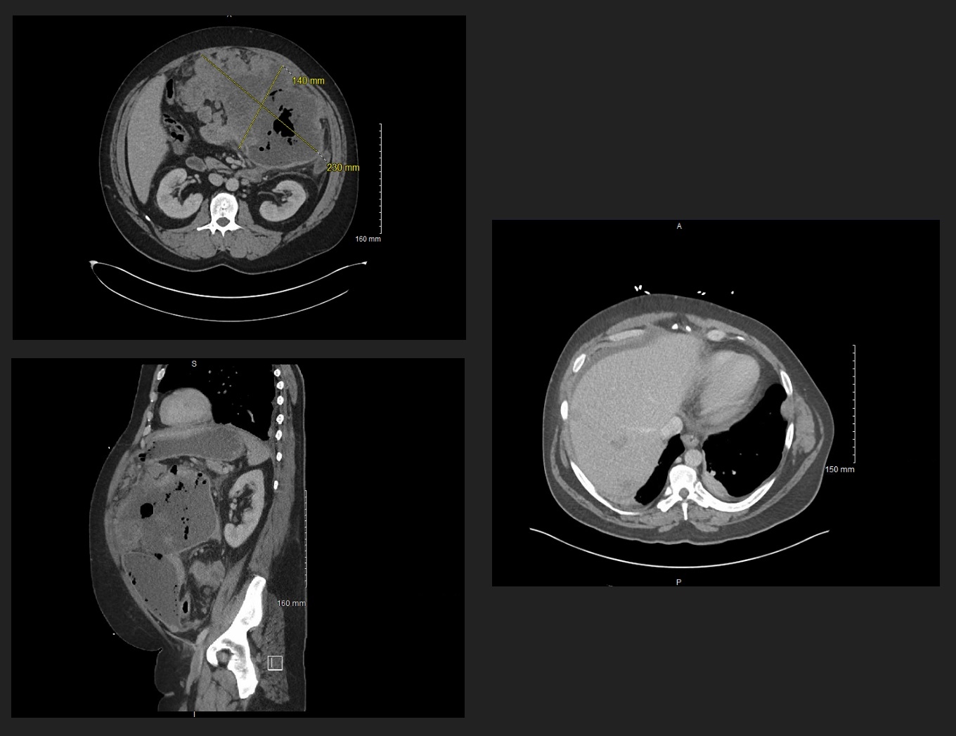
Figure: Colonoscopy images showing fungating, infiltrative mass at the splenic flexure; tattooed
Disclosures:
Mohammed Zaman indicated no relevant financial relationships.
Saad Rashid indicated no relevant financial relationships.
Fahad Sarvari indicated no relevant financial relationships.
Sultan Ahmed indicated no relevant financial relationships.
Luqman Baloch indicated no relevant financial relationships.
Altaf Dawood indicated no relevant financial relationships.
Mohammed Zaman, DO1, Saad Rashid, MD1, Fahad Sarvari, MD2, Sultan Ahmed, DO1, Luqman Baloch, MD2, Altaf Dawood, MD3. P2574 - A Case of Primary Colonic Poorly Differentiated Neuroendocrine Carcinoma With Small Cell Features, ACG 2025 Annual Scientific Meeting Abstracts. Phoenix, AZ: American College of Gastroenterology.