University of Texas Health San Antonio San Antonio, TX
Cody Hu, MD1, Hannah Yoo, MD1, Ussama Ghumman, MD1, Eugenia Tsai, MD2, Lisa D. Pedicone, PhD3, Jan Petrasek, MD, PhD3, Andres Gomez-Aldana, MD1, Eric Lawitz, MD2, Fred Poordad, MD1, Carmen Landaverde, MD3, Jason Rocha, MD1, Fabian Rodas, MD1 1University of Texas Health San Antonio, San Antonio, TX; 2Texas Liver Institute, San Antonio, TX; 3Texas Liver Institute, Austin, TX Introduction: A solitary fibrous tumor (SFT) is a rare and typically benign fibroblastic mesenchymal neoplasm arising from intrathoracic sites. It has an age-adjusted yearly incidence rate of 1 per million people and the liver is a rare site of SFT involvement. We report a case of aggressive hepatic SFT with an accompanying review of the literature.
Case Description/
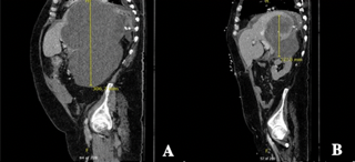
Methods: A 68-year-old man with hypertension and hyperlipidemia presented with vomiting and diarrhea. CT of the abdomen revealed hemoperitoneum and a large multiloculated hypoattenuating liver mass measuring 30 cm in diameter, causing mass effect with near collapse of the right lower lung (Figure 1A). Arterial embolization, necrosectomy, and percutaneous drainage were performed due to multiple arterial bleeds and mass effect. Blood cultures grew P. aeruginosa, E. faecalis, and K. pneumoniae during the admission. Despite appropriate antibiotics and empiric antifungal therapy, the patient clinically decompensated with interval development of abscess formation, pulmonary embolism, and persistent peritoneal bleeding. He eventually underwent open partial hepatectomy with liver cyst fenestration and surgical drain placement into the cyst cavity. Tissue pathology disclosed SFT with positive immunohistochemical (IHC) staining for CD34, vimentin, and CD99. A NAB2-STAT6 gene fusion was detected, further supporting the diagnosis of SFT. A progressively heterogeneous collection/mass was identified along the surgical bed, with both intrahepatic and extrahepatic components, raising concern for tumor aggression (Figure 1B). The patient ultimately passed despite extensive intervention. Discussion: Only 85 cases of hepatic SFT were reported in English-language literature between 1958 and 2019. Since then (Jan 2020-Oct 2024), only 10 additional primary hepatic SFT cases, including this one, have been identified per our review (Table 1), with only 2 of the 10 demonstrating aggressive behavior. SFT generally presents with nonspecific imaging characteristics, and due to its rarity, the diagnosis relies primarily on biopsy with molecular testing. STAT6 IHC serves as both a sensitive and specific marker for SFT with NAB2-STAT6 gene fusion more associated with a malignant form, as seen in our case. Although hepatic SFT is rare, the use of immunohistochemistry with STAT6 becomes relevant in tumors with rare tumor characteristics on imaging and ambiguous liver biopsy.
Figure: Figure 1. Liver mass on CT Abdomen (A) initial presentation as large multiloculated hypoattenuating liver mass measuring approximately 30cm (B) post-surgical change concerning for residual lesion and/or tumor aggression.
Figure: Table 1. Clinical data of primary hepatic solitary fibrous tumor in patients from 1/1/2020 to 10/15/2024
Cody Hu, MD1, Hannah Yoo, MD1, Ussama Ghumman, MD1, Eugenia Tsai, MD2, Lisa D. Pedicone, PhD3, Jan Petrasek, MD, PhD3, Andres Gomez-Aldana, MD1, Eric Lawitz, MD2, Fred Poordad, MD1, Carmen Landaverde, MD3, Jason Rocha, MD1, Fabian Rodas, MD1. P6100 - From Cyst to Crisis: A Giant Solitary Fibrous Tumor of the Liver, ACG 2025 Annual Scientific Meeting Abstracts. Phoenix, AZ: American College of Gastroenterology.