Rama Mouhaffel, MD1, Joseph Kim, MD1, Ramzi Ibrahim, MD2, Hoang Nhat Pham, MD3, Nazli Begum Ozturk, MD4, Samuel Cheong, DO5, Carlos Villarroel, MD1, Belinda Sun, MD, PhD1, Rolando J. Leal, MD6, Neha Jaswal, MBBS1 1Banner - University of Arizona Tucson, Tucson, AZ; 2Mayo Clinic, Scottsdale, AZ; 3Banner - University of Arizona Tucson, Phoenix, AZ; 4Corewell Health William Beaumont University Hospital, Royal Oak, MI; 5University of Arizona College of Medicine, Tucson, AZ; 6Banner University Medical Center, Tucson, AZ Introduction: Systemic amyloidosis is frequently under diagnosed as it presents with nonspecific symptoms and can affect any organ system. This case report discusses the unusual case of an asymptomatic 52-year-old male who was discovered to have elevated liver enzymes and subsequently diagnosed with AL amyloidosis as discovered by a liver biopsy.
Case Description/
Methods: A 52-year-old male with a history of CAD presented to the ED for evaluation of persistent jaundice and elevated transaminases. A RUQ ultrasound was ordered which demonstrated hepatomegaly with steatosis and concern for chronic liver disease (CLD). The patient was discharged home with follow-up with his PCP. At his follow-up, his liver function tests had further increased: AST 197, ALT 132, Alkaline phosphatase 1603, total bilirubin 4.1, and GGT 2023. The patient returned to the ED and underwent further workup, including further labs and imaging. A CT abdomen/pelvis demonstrated a decompressed gallbladder with reactive gallbladder wall edema and no significant dilation of the intrahepatic biliary ducts. An MRI of the abdomen demonstrated hepatosplenomegaly with no evidence of obstructive biliopathy. Further laboratory workup was unremarkable, including ANA, anti-smooth muscle antibody, anti-microsomal antibody, ceruloplasmin, alpha-1-antitrypsin, iron panel and hepatitis panel.
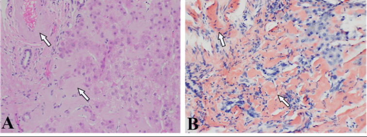
Given the patient's unexplained elevated transaminases, a trans-jugular liver biopsy was performed. Results were remarkable for apple-green birefringence with Congo red staining. Pathology was significant for severe amyloidosis deposition present in vascular wall and hepatocyte parenchyma as shown in Figure 1. Further workup was performed, including EGD and colonoscopy. Biopsies demonstrated amyloid deposition as shown in Figure 2. The patient was diagnosed with systemic amyloidosis. The patient underwent a bone marrow biopsy which supported the diagnosis of AL amyloidosis. He was subsequently started on Daratumumab for treatment. Discussion: This case focuses on the implications of amyloidosis on chronic liver disease. Patients with amyloidosis who present as chronic liver disease are frequently misdiagnosed and are delayed further care, as with our patient. Additionally, this patient was an unusual presentation of AL amyloidosis, as the patient was asymptomatic. This case reflects the importance of amyloidosis as a consideration in newly diagnosed CLD, especially in patients who do not have predisposing risk factors to CLD, such as obesity, alcohol use, or hepatitis.
Figure: Figure 1. A: Liver biopsy shows diffuse deposition of pink, glassy appearing amyloid material in the vascular wall and extracellular hepatocyte parenchyma (arrows). B: Congo Red Stain shows "salmon-pink" amyloid material (arrows) under regular light microscope and appearing apple-green birefringence under polarized light microscope.
Figure: Figure 2. A: Small intestine biopsy showing deposition of homogenous, waxy, an eosinophilic extracellular material, and its deposition in the vasculature. B: Congo-red stain light microscopy highlights the "salmon-pink" coloration of the extracellular material, which extends beyond the vessels. C: Congo-red stain under polarized light shows the characteristic "apple-green" birefringence of the amyloid material. D: Texas-Red Fluorescence Microscopy of Congo-red stained tissue enhances the visualization of amyloid deposition, especially in vessels.
Disclosures: Rama Mouhaffel indicated no relevant financial relationships. Joseph Kim indicated no relevant financial relationships. Ramzi Ibrahim indicated no relevant financial relationships. Hoang Nhat Pham indicated no relevant financial relationships. Nazli Begum Ozturk indicated no relevant financial relationships. Samuel Cheong indicated no relevant financial relationships. Carlos Villarroel indicated no relevant financial relationships. Belinda Sun indicated no relevant financial relationships. Rolando Leal indicated no relevant financial relationships. Neha Jaswal indicated no relevant financial relationships.
Rama Mouhaffel, MD1, Joseph Kim, MD1, Ramzi Ibrahim, MD2, Hoang Nhat Pham, MD3, Nazli Begum Ozturk, MD4, Samuel Cheong, DO5, Carlos Villarroel, MD1, Belinda Sun, MD, PhD1, Rolando J. Leal, MD6, Neha Jaswal, MBBS1. P6158 - Systemic Light Chain Amyloidosis Presenting as Chronic Liver Disease, ACG 2025 Annual Scientific Meeting Abstracts. Phoenix, AZ: American College of Gastroenterology.