Kavina Munshi, DO1, Tehilla Apfel, MD2 1Jefferson Torresdale Hospital, Philadelphia, PA; 2Jefferson Health, Langhorne, PA Introduction: Sclerosing mesenteritis is a rare, idiopathic fibroinflammatory disease of the mesentery. It often presents with nonspecific symptoms like abdominal pain, as seen in this case of a woman with persistent lower abdominal pain. Diagnosis is challenging, but imaging features such as a “misty mesentery” can aid in making the diagnosis. Treatment with immunosuppressants may lead to symptom relief.
Case Description/
Methods: A 49 year old female with a past medical history of chronic back pain and migraines presented with lower abdominal pain and hematochezia. She had a CT scan of her abdomen and pelvis, which showed evidence of mesenteric adenitis. She then had a colonoscopy which only showed internal hemorrhoids. Following discharge, she presented to Gastroenterology outpatient and had a repeat CT scan showing hazy attenuation of small intestinal mesentery with associated small lymph nodes, findings suggestive of sclerosing mesenteritis. She was referred to Oncology who ruled out malignancy. She then followed up with Gastroenterology due to continued lower abdominal pain with associated nausea and vomiting. Repeat imaging remained unchanged. She then had upper endoscopy to evaluate for alternative etiologies. This did not show evidence of other pathology that would contribute to her symptoms. With an absence of evidence for an alternative diagnosis, the patient’s symptoms were thought to be due to sclerosing mesenteritis. She then saw Rheumatology for workup. Her inflammatory markers were normal but she was started on methotrexate. Repeat imaging showed no evidence of inflammation and her symptoms improved with treatment. Discussion: This case highlights the complexity of diagnosing sclerosing mesenteritis, a rare fibroinflammatory disorder affecting the small bowel mesentery. The patient’s persistent abdominal pain, imaging findings, and lymphadenopathy supported this diagnosis after ruling out other causes. Although the exact etiology is unknown, there are thought to be associations with autoimmune conditions, trauma, and chronic infection. Imaging features like “misty mesentery” are typical, but definitive diagnosis requires histological confirmation. The patient responded well to immunosuppressive therapy, showing symptom relief and resolution of inflammatory changes on imaging. This case highlights the need to consider sclerosing mesenteritis in patients with unexplained mesenteric abnormalities and that treatment should be considered even in the absence of positive inflammatory markers.
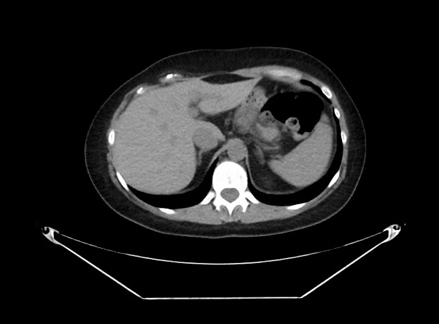
Figure: The initial CT abdomen pelvis with findings of hazy attenuation of the small intestine mesentery with associated small lymph nodes.
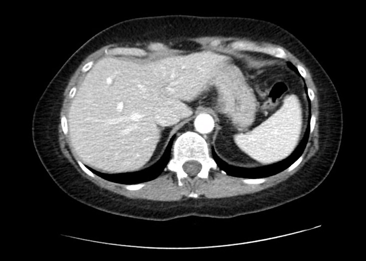
Figure: CT scan of the abdomen and pelvis without inflammatory changes seen on prior imaging studies.
Disclosures: Kavina Munshi indicated no relevant financial relationships. Tehilla Apfel indicated no relevant financial relationships.
Kavina Munshi, DO1, Tehilla Apfel, MD2. P6271 - Mesenteric Mysteries: Diagnosing and Treating the Rare Sclerosing Mesenteritis, ACG 2025 Annual Scientific Meeting Abstracts. Phoenix, AZ: American College of Gastroenterology.

 photo")