University of Arizona College of Medicine Tucson, AZ
Franklin Liu, MD1, Samuel Cheong, DO1, Joseph Gunderson, MD2, Sabrina Ho, MD1, Kai Tey, MBBS3 1University of Arizona College of Medicine, Tucson, AZ; 2University of Texas Southwestern Medical Center, Dallas, TX; 3Banner - University of Arizona Tucson, Tucson, AZ Introduction: Hepatic hemangiomas are the most common benign liver tumors, typically asymptomatic and discovered incidentally on imaging. Surgical intervention is indicated in fewer than 1% of cases and is generally reserved for patients with complications such as rupture, hemorrhage, consumptive coagulopathy, or significant compression of adjacent organs. Although most hepatic hemangiomas remain stable or decrease in size over time, “giant” hemangiomas (lesions >10 cm) account for fewer than 10% of all cases and may warrant surgery if symptoms or complications develop. Herein, we present a rare case of a giant hepatic hemangioma exhibiting progressive growth and late-onset symptoms over 13 years requiring surgical resection in an elderly female.
Case Description/
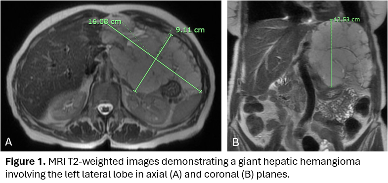
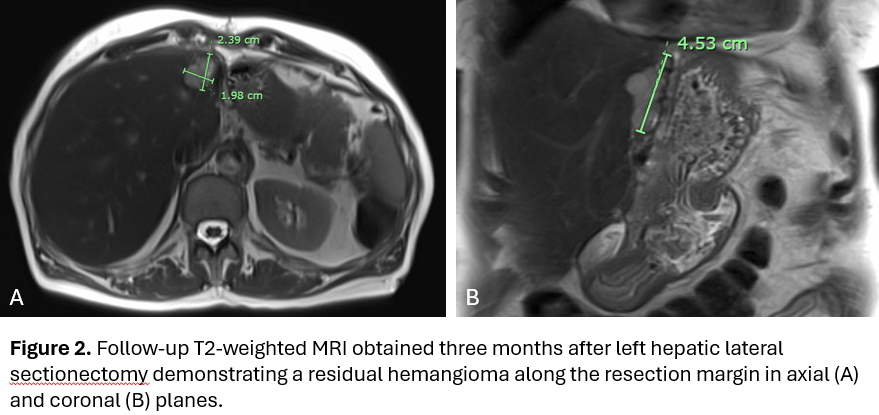
Methods: A 59-year-old woman with a history of hyperlipidemia and remote tobacco use was incidentally found to have a 10-cm hepatic hemangioma on imaging in 2010. She was asymptomatic at the time. Interval imaging in 2023 demonstrated enlargement of the lesion to 14 × 9.1 × 12.4 cm. Although she remained asymptomatic initially, by early 2024, at the age of 74, she developed progressive abdominal discomfort and distention. Abdominal magnetic resonance imaging (MRI) revealed further growth of the lesion to 16.1 × 9.1 × 12.6 cm (Figure 1). Laboratory studies, including liver function tests and platelet counts, remained normal throughout. Given the lesion’s rapid growth of over 2 cm/year and the onset of symptoms, her case was reviewed at a multidisciplinary tumor board. Surgical resection was recommended, and she underwent a left lateral sectionectomy in November 2024. Pathology confirmed a benign cavernous hemangioma measuring 16 cm. The patient had an uneventful postoperative recovery and was asymptomatic at follow-up. A surveillance MRI in February 2025 showed a small residual hemangioma at the resection margin measuring 2.4 × 2.0 × 4.5 cm Figure 2). Discussion: This case illustrates the rare but recognized occurrence of a giant hepatic hemangioma exhibiting progressive growth and late-onset symptoms requiring surgical resection in an elderly patient. Although less than 1% of hepatic hemangiomas require resection, this case illustrates that even longstanding, asymptomatic hepatic hemangiomas may warrant resection when interval growth and symptoms develop. Future work is needed to assess long-term post-operative outcomes and surveillance strategies.
Figure: Figure 1. MRI T2-weighted images demonstrating a giant hepatic hemangioma involving the left lateral lobe in axial (A) and coronal (B) planes.
Figure: Figure 2. Follow-up T2-weighted MRI obtained three months after left hepatic lateral sectionectomy demonstrating a residual hemangioma along the resection margin in axial (A) and coronal (B) planes.
Disclosures: Franklin Liu indicated no relevant financial relationships. Samuel Cheong indicated no relevant financial relationships. Joseph Gunderson indicated no relevant financial relationships. Sabrina Ho indicated no relevant financial relationships. Kai Tey indicated no relevant financial relationships.
Franklin Liu, MD1, Samuel Cheong, DO1, Joseph Gunderson, MD2, Sabrina Ho, MD1, Kai Tey, MBBS3. P6128 - Surgical Resection of a Giant Hepatic Hemangioma Following Rapid Growth and Symptom Development, ACG 2025 Annual Scientific Meeting Abstracts. Phoenix, AZ: American College of Gastroenterology.